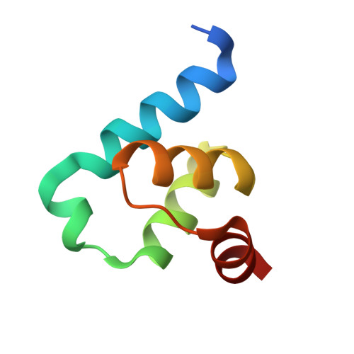
6RT2

Crystal structure of Trypanosoma Brucei PEX14 N-terminal domain in complex with small molecules designed to investigate the water envelope


Experimental Data Snapshot

  • Method: X-RAY DIFFRACTION
  • Resolution: 1.30 Å
  • R-Value Free: 0.189 
  • R-Value Work: 0.161 
  • R-Value Observed: 0.163 

wwPDB Validation   3D Report Full Report


Ligand Structure Quality Assessment 


This is version 1.2 of the entry. See complete history


Literature

Water envelope has a critical impact on the design of protein-protein interaction inhibitors.

Ratkova, E.L.Dawidowski, M.Napolitano, V.Dubin, G.Fino, R.Ostertag, M.S.Sattler, M.Popowicz, G.Tetko, I.V.

(2020) Chem Commun (Camb) 56: 4360-4363

  • DOI: https://doi.org/10.1039/c9cc07714f
  • Primary Citation of Related Structures:  
    6RT2

  • PubMed Abstract: 

    We show that a water envelope network plays a critical role in protein-protein interactions (PPI). The potency of a PPI inhibitor is modulated by orders of magnitude on manipulation of the solvent envelope alone. The structure-activity relationship of PEX14 inhibitors was analyzed as an example using in silico and X-ray data.


  • Organizational Affiliation

    Institute of Structural Biology, Helmholtz Zentrum München - German Research Center for Environmental Health (GmbH), Ingolstädter Landstraße 1, 85764 Neuherberg, Germany. grzegorz.popowicz@helmholtz-muenchen.de i.tetko@helmholtz-muenchen.de and Medicinal Chemistry, Cardiovascular, Renal and Metabolic Diseases, IMED Biotech Unit, AstraZeneca, Gothenburg, Sweden. ekaterina.ratkova@astrazeneca.com.


Macromolecules
Find similar proteins by:  (by identity cutoff)  |  3D Structure
Entity ID: 1
MoleculeChains Sequence LengthOrganismDetailsImage
Peroxin 14
A, B, C, D
67Trypanosoma brucei bruceiMutation(s): 0 
Gene Names: PEX14
UniProt
Find proteins for Q8IEW2 (Trypanosoma brucei brucei)
Explore Q8IEW2 
Go to UniProtKB:  Q8IEW2
Entity Groups  
Sequence Clusters30% Identity50% Identity70% Identity90% Identity95% Identity100% Identity
UniProt GroupQ8IEW2
Sequence Annotations
Expand
  • Reference Sequence
Small Molecules
Ligands 3 Unique
IDChains Name / Formula / InChI Key2D Diagram3D Interactions
CV5 (Subject of Investigation/LOI)
Query on CV5

Download Ideal Coordinates CCD File 
E [auth A],
H [auth B],
M [auth C],
O [auth D]
(3~{S})-3-[[1-(2-hydroxyethyl)-5-[(4-methoxynaphthalen-1-yl)methyl]-6,7-dihydro-4~{H}-pyrazolo[4,3-c]pyridin-3-yl]carbonylamino]-3-phenyl-propanoic acid
C30 H32 N4 O5
FEINFPMXJVPOSJ-VWLOTQADSA-N
SO4
Query on SO4

Download Ideal Coordinates CCD File 
F [auth A]
I [auth B]
J [auth B]
K [auth B]
P [auth D]
F [auth A],
I [auth B],
J [auth B],
K [auth B],
P [auth D],
Q [auth D]
SULFATE ION
O4 S
QAOWNCQODCNURD-UHFFFAOYSA-L
BME
Query on BME

Download Ideal Coordinates CCD File 
G [auth A],
L [auth B],
N [auth C],
R [auth D]
BETA-MERCAPTOETHANOL
C2 H6 O S
DGVVWUTYPXICAM-UHFFFAOYSA-N
Binding Affinity Annotations 
IDSourceBinding Affinity
CV5 Binding MOAD:  6RT2 Kd: 6.46e+5 (nM) from 1 assay(s)
Experimental Data & Validation

Experimental Data

  • Method: X-RAY DIFFRACTION
  • Resolution: 1.30 Å
  • R-Value Free: 0.189 
  • R-Value Work: 0.161 
  • R-Value Observed: 0.163 
  • Space Group: P 1 21 1
Unit Cell:
Length ( Å )Angle ( ˚ )
a = 35.78α = 90
b = 115.52β = 101.39
c = 38.87γ = 90
Software Package:
Software NamePurpose
XSCALEdata scaling
REFMACrefinement
PDB_EXTRACTdata extraction
XDSdata reduction
PHASERphasing

Structure Validation

View Full Validation Report


Ligand Structure Quality Assessment 


Entry History & Funding Information

Deposition Data


Funding OrganizationLocationGrant Number
Accelerated Early staGe drug dIScoveryPoland675555

Revision History  (Full details and data files)

  • Version 1.0: 2020-04-08
    Type: Initial release
  • Version 1.1: 2020-04-29
    Changes: Database references
  • Version 1.2: 2024-01-24
    Changes: Data collection, Database references, Refinement description