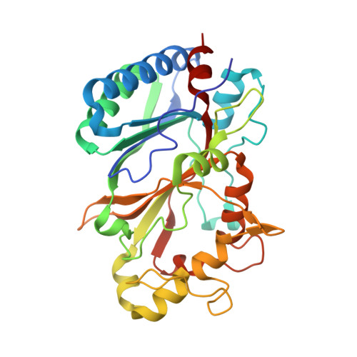
6RR6

Structure of 100% reduced KpDyP


Experimental Data Snapshot

  • Method: X-RAY DIFFRACTION
  • Resolution: 1.90 Å
  • R-Value Free: 0.199 
  • R-Value Work: 0.181 
  • R-Value Observed: 0.182 

wwPDB Validation   3D Report Full Report


Ligand Structure Quality Assessment 


This is version 1.2 of the entry. See complete history


Literature

X-ray-induced photoreduction of heme metal centers rapidly induces active-site perturbations in a protein-independent manner.

Pfanzagl, V.Beale, J.H.Michlits, H.Schmidt, D.Gabler, T.Obinger, C.Djinovic-Carugo, K.Hofbauer, S.

(2020) J Biol Chem 295: 13488-13501

  • DOI: https://doi.org/10.1074/jbc.RA120.014087
  • Primary Citation of Related Structures:  
    6RPD, 6RPE, 6RQY, 6RR1, 6RR4, 6RR5, 6RR6, 6RR8

  • PubMed Abstract: 

    Since the advent of protein crystallography, atomic-level macromolecular structures have provided a basis to understand biological function. Enzymologists use detailed structural insights on ligand coordination, interatomic distances, and positioning of catalytic amino acids to rationalize the underlying electronic reaction mechanisms. Often the proteins in question catalyze redox reactions using metal cofactors that are explicitly intertwined with their function. In these cases, the exact nature of the coordination sphere and the oxidation state of the metal is of utmost importance. Unfortunately, the redox-active nature of metal cofactors makes them especially susceptible to photoreduction, meaning that information obtained by photoreducing X-ray sources about the environment of the cofactor is the least trustworthy part of the structure. In this work we directly compare the kinetics of photoreduction of six different heme protein crystal species by X-ray radiation. We show that a dose of ∼40 kilograys already yields 50% ferrous iron in a heme protein crystal. We also demonstrate that the kinetics of photoreduction are completely independent from variables unique to the different samples tested. The photoreduction-induced structural rearrangements around the metal cofactors have to be considered when biochemical data of ferric proteins are rationalized by constraints derived from crystal structures of reduced enzymes.


  • Organizational Affiliation

    Department of Chemistry, Institute of Biochemistry, BOKU-University of Natural Resources and Life Sciences, Vienna, Austria. Electronic address: vera.pfanzagl@boku.ac.at.


Macromolecules
Find similar proteins by:  (by identity cutoff)  |  3D Structure
Entity ID: 1
MoleculeChains Sequence LengthOrganismDetailsImage
Iron-dependent peroxidase
A, B
303Klebsiella pneumoniaeMutation(s): 0 
Gene Names: 
EC: 1.11.1
UniProt
Find proteins for A0A0W8ATM9 (Klebsiella pneumoniae)
Explore A0A0W8ATM9 
Go to UniProtKB:  A0A0W8ATM9
Entity Groups  
Sequence Clusters30% Identity50% Identity70% Identity90% Identity95% Identity100% Identity
UniProt GroupA0A0W8ATM9
Sequence Annotations
Expand
  • Reference Sequence
Small Molecules
Ligands 3 Unique
IDChains Name / Formula / InChI Key2D Diagram3D Interactions
HEM (Subject of Investigation/LOI)
Query on HEM

Download Ideal Coordinates CCD File 
C [auth A],
K [auth B]
PROTOPORPHYRIN IX CONTAINING FE
C34 H32 Fe N4 O4
KABFMIBPWCXCRK-RGGAHWMASA-L
GOL
Query on GOL

Download Ideal Coordinates CCD File 
D [auth A]
E [auth A]
F [auth A]
G [auth A]
H [auth A]
D [auth A],
E [auth A],
F [auth A],
G [auth A],
H [auth A],
I [auth A],
L [auth B],
M [auth B],
N [auth B],
O [auth B]
GLYCEROL
C3 H8 O3
PEDCQBHIVMGVHV-UHFFFAOYSA-N
MG
Query on MG

Download Ideal Coordinates CCD File 
J [auth A],
P [auth B]
MAGNESIUM ION
Mg
JLVVSXFLKOJNIY-UHFFFAOYSA-N
Experimental Data & Validation

Experimental Data

  • Method: X-RAY DIFFRACTION
  • Resolution: 1.90 Å
  • R-Value Free: 0.199 
  • R-Value Work: 0.181 
  • R-Value Observed: 0.182 
  • Space Group: P 1 21 1
Unit Cell:
Length ( Å )Angle ( ˚ )
a = 50.889α = 90
b = 76.647β = 108.01
c = 76.579γ = 90
Software Package:
Software NamePurpose
BUSTERrefinement
XDSdata reduction
Aimlessdata scaling
PHENIXphasing

Structure Validation

View Full Validation Report


Ligand Structure Quality Assessment 


Entry History 

Deposition Data

Revision History  (Full details and data files)

  • Version 1.0: 2020-09-02
    Type: Initial release
  • Version 1.1: 2020-10-07
    Changes: Database references
  • Version 1.2: 2024-01-24
    Changes: Data collection, Database references, Refinement description