6RK4

Lysostaphin SH3b P4-G5 complex, synchrotron dataset


Experimental Data Snapshot

  • Method: X-RAY DIFFRACTION
  • Resolution: 1.43 Å
  • R-Value Free: 0.230 
  • R-Value Work: 0.199 
  • R-Value Observed: 0.200 

wwPDB Validation   3D Report Full Report


Ligand Structure Quality Assessment 


This is version 1.3 of the entry. See complete history


Literature

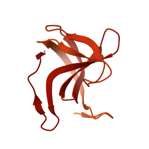
Two-site recognition of Staphylococcus aureus peptidoglycan by lysostaphin SH3b.

Gonzalez-Delgado, L.S.Walters-Morgan, H.Salamaga, B.Robertson, A.J.Hounslow, A.M.Jagielska, E.Sabala, I.Williamson, M.P.Lovering, A.L.Mesnage, S.

(2020) Nat Chem Biol 16: 24-30

  • DOI: https://doi.org/10.1038/s41589-019-0393-4
  • Primary Citation of Related Structures:  
    6RJE, 6RK4

  • PubMed Abstract: 

    Lysostaphin is a bacteriolytic enzyme targeting peptidoglycan, the essential component of the bacterial cell envelope. It displays a very potent and specific activity toward staphylococci, including methicillin-resistant Staphylococcus aureus. Lysostaphin causes rapid cell lysis and disrupts biofilms, and is therefore a therapeutic agent of choice to eradicate staphylococcal infections. The C-terminal SH3b domain of lysostaphin recognizes peptidoglycans containing a pentaglycine crossbridge and has been proposed to drive the preferential digestion of staphylococcal cell walls. Here we elucidate the molecular mechanism underpinning recognition of staphylococcal peptidoglycan by the lysostaphin SH3b domain. We show that the pentaglycine crossbridge and the peptide stem are recognized by two independent binding sites located on opposite sides of the SH3b domain, thereby inducing a clustering of SH3b domains. We propose that this unusual binding mechanism allows synergistic and structurally dynamic recognition of S. aureus peptidoglycan and underpins the potent bacteriolytic activity of this enzyme.


  • Organizational Affiliation

    Department of Molecular Biology and Biotechnology, University of Sheffield, Sheffield, UK.


Macromolecules
Find similar proteins by:  (by identity cutoff)  |  3D Structure
Entity ID: 1
MoleculeChains Sequence LengthOrganismDetailsImage
Lysostaphin493Staphylococcus simulansMutation(s): 0 
Gene Names: lss
EC: 3.4.24.75
UniProt
Find proteins for P10547 (Staphylococcus simulans)
Explore P10547 
Go to UniProtKB:  P10547
Entity Groups  
Sequence Clusters30% Identity50% Identity70% Identity90% Identity95% Identity100% Identity
UniProt GroupP10547
Sequence Annotations
Expand
  • Reference Sequence
Small Molecules
Ligands 2 Unique
IDChains Name / Formula / InChI Key2D Diagram3D Interactions
K5T (Subject of Investigation/LOI)
Query on K5T

Download Ideal Coordinates CCD File 
B [auth A](2~{R})-2-[[(2~{S})-2-[[(4~{R})-5-azanyl-4-[[(2~{S})-2-azanylpropanoyl]amino]-5-oxidanylidene-pentanoyl]amino]-6-[2-[2-[2-[2-(2-azanylethanoylamino)ethanoylamino]ethanoylamino]ethanoylamino]ethanoylamino]hexanoyl]amino]propanoic acid
C27 H47 N11 O11
HUYIRRKHPBSVEV-HZMVEIRTSA-N
EDO
Query on EDO

Download Ideal Coordinates CCD File 
C [auth A],
D [auth A]
1,2-ETHANEDIOL
C2 H6 O2
LYCAIKOWRPUZTN-UHFFFAOYSA-N
Binding Affinity Annotations 
IDSourceBinding Affinity
K5T Binding MOAD:  6RK4 Kd: 8.90e+5 (nM) from 1 assay(s)
Experimental Data & Validation

Experimental Data

  • Method: X-RAY DIFFRACTION
  • Resolution: 1.43 Å
  • R-Value Free: 0.230 
  • R-Value Work: 0.199 
  • R-Value Observed: 0.200 
  • Space Group: P 41 21 2
Unit Cell:
Length ( Å )Angle ( ˚ )
a = 47.08α = 90
b = 47.08β = 90
c = 122.35γ = 90
Software Package:
Software NamePurpose
XDSdata reduction
Aimlessdata scaling
PHASERphasing
REFMACrefinement
PDB_EXTRACTdata extraction

Structure Validation

View Full Validation Report


Ligand Structure Quality Assessment 


Entry History & Funding Information

Deposition Data


Funding OrganizationLocationGrant Number
Biotechnology and Biological Sciences Research CouncilUnited KingdomBB/R000727/1
Engineering and Physical Sciences Research CouncilUnited KingdomEP/S01358X/1
Biotechnology and Biological Sciences Research CouncilUnited KingdomMIBTP studentship

Revision History  (Full details and data files)

  • Version 1.0: 2019-10-16
    Type: Initial release
  • Version 1.1: 2019-11-20
    Changes: Database references
  • Version 1.2: 2019-12-25
    Changes: Database references
  • Version 1.3: 2024-01-24
    Changes: Data collection, Database references, Refinement description