6R3M

Family 11 Carbohydrate-Binding Module from Clostridium thermocellum in complex with beta-1,3-1,4-mixed-linked tetrasaccharide


Experimental Data Snapshot

  • Method: X-RAY DIFFRACTION
  • Resolution: 1.45 Å
  • R-Value Free: 0.208 
  • R-Value Work: 0.177 
  • R-Value Observed: 0.179 

wwPDB Validation   3D Report Full Report


Ligand Structure Quality Assessment 


This is version 2.1 of the entry. See complete history


Literature

Molecular basis for the preferential recognition of beta 1,3-1,4-glucans by the family 11 carbohydrate-binding module from Clostridium thermocellum.

Ribeiro, D.O.Viegas, A.Pires, V.M.R.Medeiros-Silva, J.Bule, P.Chai, W.Marcelo, F.Fontes, C.M.G.A.Cabrita, E.J.Palma, A.S.Carvalho, A.L.

(2020) FEBS J 287: 2723-2743

  • DOI: https://doi.org/10.1111/febs.15162
  • Primary Citation of Related Structures:  
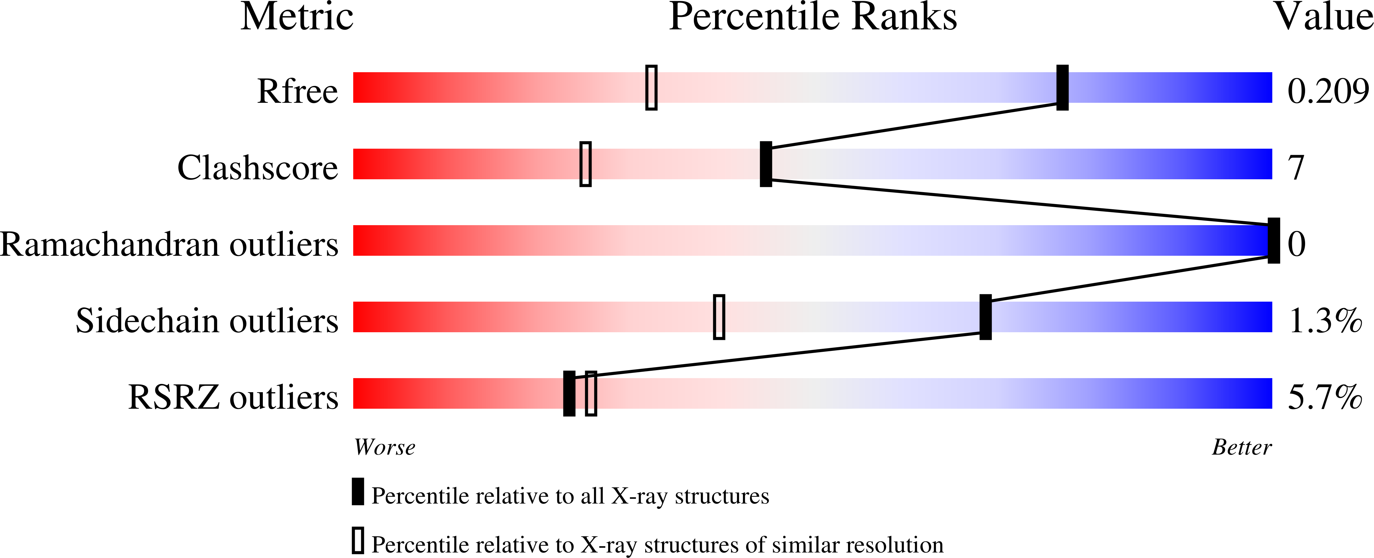
    6R31, 6R3M

  • PubMed Abstract: 

    Understanding the specific molecular interactions between proteins and β1,3-1,4-mixed-linked d-glucans is fundamental to harvest the full biological and biotechnological potential of these carbohydrates and of proteins that specifically recognize them. The family 11 carbohydrate-binding module from Clostridium thermocellum (CtCBM11) is known for its binding preference for β1,3-1,4-mixed-linked over β1,4-linked glucans. Despite the growing industrial interest of this protein for the biotransformation of lignocellulosic biomass, the molecular determinants of its ligand specificity are not well defined. In this report, a combined approach of methodologies was used to unravel, at a molecular level, the ligand recognition of CtCBM11. The analysis of the interaction by carbohydrate microarrays and NMR and the crystal structures of CtCBM11 bound to β1,3-1,4-linked glucose oligosaccharides showed that both the chain length and the position of the β1,3-linkage are important for recognition, and identified the tetrasaccharide Glcβ1,4Glcβ1,4Glcβ1,3Glc sequence as a minimum epitope required for binding. The structural data, along with site-directed mutagenesis and ITC studies, demonstrated the specificity of CtCBM11 for the twisted conformation of β1,3-1,4-mixed-linked glucans. This is mediated by a conformation-selection mechanism of the ligand in the binding cleft through CH-π stacking and a hydrogen bonding network, which is dependent not only on ligand chain length, but also on the presence of a β1,3-linkage at the reducing end and at specific positions along the β1,4-linked glucan chain. The understanding of the detailed mechanism by which CtCBM11 can distinguish between linear and mixed-linked β-glucans strengthens its exploitation for the design of new biomolecules with improved capabilities and applications in health and agriculture. DATABASE: Structural data are available in the Protein Data Bank under the accession codes 6R3M and 6R31.


  • Organizational Affiliation

    UCIBIO, Departamento de Química, Faculdade de Ciências e Tecnologia, Universidade NOVA de Lisboa, Caparica, Portugal.


Macromolecules
Find similar proteins by:  (by identity cutoff)  |  3D Structure
Entity ID: 1
MoleculeChains Sequence LengthOrganismDetailsImage
Endoglucanase H178Acetivibrio thermocellus ATCC 27405Mutation(s): 0 
Gene Names: celHCthe_1472
EC: 3.2.1.4
UniProt
Find proteins for P16218 (Acetivibrio thermocellus (strain ATCC 27405 / DSM 1237 / JCM 9322 / NBRC 103400 / NCIMB 10682 / NRRL B-4536 / VPI 7372))
Explore P16218 
Go to UniProtKB:  P16218
Entity Groups  
Sequence Clusters30% Identity50% Identity70% Identity90% Identity95% Identity100% Identity
UniProt GroupP16218
Sequence Annotations
Expand
  • Reference Sequence
Oligosaccharides

Help

Entity ID: 2
MoleculeChains Length2D Diagram Glycosylation3D Interactions
beta-D-glucopyranose-(1-4)-beta-D-glucopyranose-(1-4)-beta-D-glucopyranose-(1-3)-beta-D-glucopyranose
B
4N/A
Glycosylation Resources
GlyTouCan:  G95849GT
GlyCosmos:  G95849GT
GlyGen:  G95849GT
Small Molecules
Ligands 4 Unique
IDChains Name / Formula / InChI Key2D Diagram3D Interactions
GLC
Query on GLC

Download Ideal Coordinates CCD File 
J [auth A]alpha-D-glucopyranose
C6 H12 O6
WQZGKKKJIJFFOK-DVKNGEFBSA-N
PO4
Query on PO4

Download Ideal Coordinates CCD File 
F [auth A],
G [auth A],
H [auth A],
I [auth A]
PHOSPHATE ION
O4 P
NBIIXXVUZAFLBC-UHFFFAOYSA-K
ACT
Query on ACT

Download Ideal Coordinates CCD File 
E [auth A]ACETATE ION
C2 H3 O2
QTBSBXVTEAMEQO-UHFFFAOYSA-M
CA
Query on CA

Download Ideal Coordinates CCD File 
C [auth A],
D [auth A]
CALCIUM ION
Ca
BHPQYMZQTOCNFJ-UHFFFAOYSA-N
Experimental Data & Validation

Experimental Data

  • Method: X-RAY DIFFRACTION
  • Resolution: 1.45 Å
  • R-Value Free: 0.208 
  • R-Value Work: 0.177 
  • R-Value Observed: 0.179 
  • Space Group: H 3
Unit Cell:
Length ( Å )Angle ( ˚ )
a = 103.249α = 90
b = 103.249β = 90
c = 39.58γ = 120
Software Package:
Software NamePurpose
REFMACrefinement
MOSFLMdata reduction
SCALAdata scaling
PHASERphasing

Structure Validation

View Full Validation Report


Ligand Structure Quality Assessment 


Entry History & Funding Information

Deposition Data


Funding OrganizationLocationGrant Number
Foundation for Science and TechnologyPortugalSFRH/BD/100569/2014
Foundation for Science and TechnologyPortugalPTDC/BBB-BEP/0869/2014
Foundation for Science and TechnologyPortugalEXPL/IF/01621/2013

Revision History  (Full details and data files)

  • Version 1.0: 2020-02-05
    Type: Initial release
  • Version 1.1: 2020-07-15
    Changes: Database references
  • Version 2.0: 2020-07-29
    Type: Remediation
    Reason: Carbohydrate remediation
    Changes: Atomic model, Data collection, Derived calculations, Structure summary
  • Version 2.1: 2024-01-24
    Changes: Data collection, Database references, Refinement description, Structure summary