6QWI

Structure of beta-glucosidase A from Paenibacillus polymyxa complexed with multivalent inhibitors.


Experimental Data Snapshot

  • Method: X-RAY DIFFRACTION
  • Resolution: 2.85 Å
  • R-Value Free: 0.231 
  • R-Value Work: 0.181 
  • R-Value Observed: 0.183 

wwPDB Validation   3D Report Full Report


Ligand Structure Quality Assessment 


This is version 1.1 of the entry. See complete history


Literature

Structural basis of the inhibition of GH1 beta-glucosidases by multivalent pyrrolidine iminosugars.

Martinez-Bailen, M.Jimenez-Ortega, E.Carmona, A.T.Robina, I.Sanz-Aparicio, J.Talens-Perales, D.Polaina, J.Matassini, C.Cardona, F.Moreno-Vargas, A.J.

(2019) Bioorg Chem 89: 103026-103026

  • DOI: https://doi.org/10.1016/j.bioorg.2019.103026
  • Primary Citation of Related Structures:  
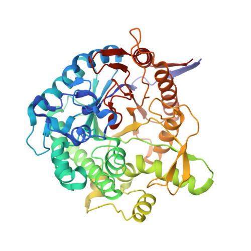
    6QWI, 6R4K

  • PubMed Abstract: 

    The synthesis of multivalent pyrrolidine iminosugars via CuAAC click reaction between different pyrrolidine-azide derivatives and tri- or hexavalent alkynyl scaffolds is reported. The new multimeric compounds, together with the monomeric reference, were evaluated as inhibitors against two homologous GH1 β-glucosidases (BglA and BglB from Paenibacillus polymyxa). The multivalent inhibitors containing an aromatic moiety in the linker between the pyrrolidine and the scaffold inhibited the octameric BglA (µM range) but did not show affinity against the monomeric BglB, despite the similarity between the active site of both enzymes. A modest multivalent effect (rp/n = 12) was detected for the hexavalent inhibitor 12. Structural analysis of the complexes between the monomeric and the trimeric iminosugar inhibitors (4 and 10) and BglA showed the insertion of the inhibitors at the active site of BglA, confirming a competitive mode of inhibition as indicated by enzyme kinetics. Additionally, structural comparison of the BglA/4 complex with the reported BglB/2F-glucose complex illustrates the key determinants responsible for the inhibitory effect and explains the reasons of the inhibition of BglA and the no inhibition of BglB. Potential inhibition of other β-glucosidases with therapeutic relevance is discussed under the light of these observations.


  • Organizational Affiliation

    Department of Organic Chemistry, Faculty of Chemistry, University of Seville, C/Prof. García González, 1, 41012 Seville, Spain.


Macromolecules
Find similar proteins by:  (by identity cutoff)  |  3D Structure
Entity ID: 1
MoleculeChains Sequence LengthOrganismDetailsImage
Beta-glucosidase A
A, B
448Paenibacillus polymyxaMutation(s): 0 
Gene Names: bglA
EC: 3.2.1.21
UniProt
Find proteins for P22073 (Paenibacillus polymyxa)
Explore P22073 
Go to UniProtKB:  P22073
Entity Groups  
Sequence Clusters30% Identity50% Identity70% Identity90% Identity95% Identity100% Identity
UniProt GroupP22073
Sequence Annotations
Expand
  • Reference Sequence
Small Molecules
Ligands 1 Unique
IDChains Name / Formula / InChI Key2D Diagram3D Interactions
JJW
Query on JJW

Download Ideal Coordinates CCD File 
C [auth A],
D [auth B]
(2~{S},3~{S},4~{R})-2-[[4-[4-(2-ethoxyethoxy)phenyl]-1,2,3-triazol-1-yl]methyl]pyrrolidine-3,4-diol
C17 H24 N4 O4
NXQXJVDZMOHAEW-BBWFWOEESA-N
Experimental Data & Validation

Experimental Data

  • Method: X-RAY DIFFRACTION
  • Resolution: 2.85 Å
  • R-Value Free: 0.231 
  • R-Value Work: 0.181 
  • R-Value Observed: 0.183 
  • Space Group: P 4 21 2
Unit Cell:
Length ( Å )Angle ( ˚ )
a = 146.096α = 90
b = 146.096β = 90
c = 140.345γ = 90
Software Package:
Software NamePurpose
REFMACrefinement
XDSdata reduction
Aimlessdata scaling
MOLREPphasing

Structure Validation

View Full Validation Report


Ligand Structure Quality Assessment 


Entry History 

Deposition Data

Revision History  (Full details and data files)

  • Version 1.0: 2019-07-03
    Type: Initial release
  • Version 1.1: 2024-01-24
    Changes: Data collection, Database references, Refinement description