The basis for non-canonical ROK family function in theN-acetylmannosamine kinase from the pathogenStaphylococcus aureus.
Coombes, D., Davies, J.S., Newton-Vesty, M.C., Horne, C.R., Setty, T.G., Subramanian, R., Moir, J.W.B., Friemann, R., Panjikar, S., Griffin, M.D.W., North, R.A., Dobson, R.C.J.(2020) J Biol Chem 295: 3301-3315
- PubMed: 31949045
- DOI: https://doi.org/10.1074/jbc.RA119.010526
- Primary Citation of Related Structures:
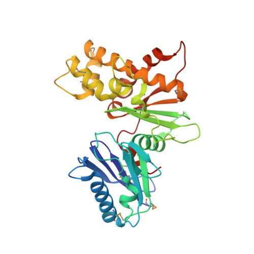
6Q26, 6Q27, 6Q28 - PubMed Abstract:
In environments where glucose is limited, some pathogenic bacteria metabolize host-derived sialic acid as a nutrient source. N -Acetylmannosamine kinase (NanK) is the second enzyme of the bacterial sialic acid import and degradation pathway and adds phosphate to N -acetylmannosamine using ATP to prime the molecule for future pathway reactions. Sequence alignments reveal that Gram-positive NanK enzymes belong to the Repressor, ORF, Kinase (ROK) family, but many lack the canonical Zn-binding motif expected for this function, and the sugar-binding E X GH motif is altered to E X GY. As a result, it is unclear how they perform this important reaction. Here, we study the Staphylococcus aureus NanK ( Sa NanK), which is the first characterization of a Gram-positive NanK. We report the kinetic activity of Sa NanK along with the ligand-free, N -acetylmannosamine-bound and substrate analog GlcNAc-bound crystal structures (2.33, 2.20, and 2.20 Å resolution, respectively). These demonstrate, in combination with small-angle X-ray scattering, that Sa NanK is a dimer that adopts a closed conformation upon substrate binding. Analysis of the E X GY motif reveals that the tyrosine binds to the N -acetyl group to select for the "boat" conformation of N -acetylmannosamine. Moreover, Sa NanK has a stacked arginine pair coordinated by negative residues critical for thermal stability and catalysis. These combined elements serve to constrain the active site and orient the substrate in lieu of Zn binding, representing a significant departure from canonical NanK binding. This characterization provides insight into differences in the ROK family and highlights a novel area for antimicrobial discovery to fight Gram-positive and S. aureus infections.
Organizational Affiliation:
Biomolecular Interaction Centre and School of Biological Sciences, University of Canterbury, Christchurch 8140, New Zealand.