6PTY

Soluble model of human CuA (Tt3Lh)


Experimental Data Snapshot

  • Method: X-RAY DIFFRACTION
  • Resolution: 1.98 Å
  • R-Value Free: 0.238 
  • R-Value Work: 0.198 
  • R-Value Observed: 0.200 

wwPDB Validation   3D Report Full Report


This is version 1.3 of the entry. See complete history


Literature

Unexpected electron spin density on the axial methionine ligand in CuAsuggests its involvement in electron pathways.

Morgada, M.N.Llases, M.E.Giannini, E.Castro, M.A.Alzari, P.M.Murgida, D.H.Lisa, M.N.Vila, A.J.

(2020) Chem Commun (Camb) 56: 1223-1226

  • DOI: https://doi.org/10.1039/c9cc08883k
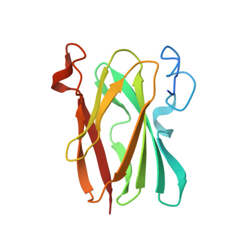
  • Primary Citation of Related Structures:  
    6PTT, 6PTY

  • PubMed Abstract: 

    The CuA center is a paradigm for the study of long-range biological electron transfer. This metal center is an essential cofactor for terminal oxidases like cytochrome c oxidase, the enzymatic complex responsible for cellular respiration in eukaryotes and in most bacteria. CuA acts as an electron hub by transferring electrons from reduced cytochrome c to the catalytic site of the enzyme where dioxygen reduction takes place. Different electron transfer pathways have been proposed involving a weak axial methionine ligand residue, conserved in all CuA sites. This hypothesis has been challenged by theoretical calculations indicating the lack of electron spin density in this ligand. Here we report an NMR study with selectively labeled methionine in a native CuA. NMR spectroscopy discloses the presence of net electron spin density in the methionine axial ligand in the two alternative ground states of this metal center. Similar spin delocalization observed on two second sphere mutants further supports this evidence. These data provide a novel view of the electronic structure of CuA centers and support previously neglected electron transfer pathways.


  • Organizational Affiliation

    Instituto de Biología Molecular y Celular de Rosario (IBR, CONICET-UNR), Ocampo y Esmeralda, Rosario 2000, Argentina. vila@ibr-conicet.gov.ar.


Macromolecules
Find similar proteins by:  (by identity cutoff)  |  3D Structure
Entity ID: 1
MoleculeChains Sequence LengthOrganismDetailsImage
Cytochrome c oxidase subunit 2
A, B
129Thermus thermophilusMutation(s): 0 
Gene Names: cbaBctaC
EC: 1.9.3.1
UniProt
Find proteins for Q5SJ80 (Thermus thermophilus (strain ATCC 27634 / DSM 579 / HB8))
Explore Q5SJ80 
Go to UniProtKB:  Q5SJ80
Entity Groups  
Sequence Clusters30% Identity50% Identity70% Identity90% Identity95% Identity100% Identity
UniProt GroupQ5SJ80
Sequence Annotations
Expand
  • Reference Sequence
Experimental Data & Validation

Experimental Data

  • Method: X-RAY DIFFRACTION
  • Resolution: 1.98 Å
  • R-Value Free: 0.238 
  • R-Value Work: 0.198 
  • R-Value Observed: 0.200 
  • Space Group: P 21 21 21
Unit Cell:
Length ( Å )Angle ( ˚ )
a = 51.63α = 90
b = 70.957β = 90
c = 77.415γ = 90
Software Package:
Software NamePurpose
PHENIXrefinement
XDSdata reduction
Aimlessdata scaling
PHASERphasing

Structure Validation

View Full Validation Report


Entry History & Funding Information

Deposition Data


Funding OrganizationLocationGrant Number
National Research Council (NRC, Argentina)ArgentinaPICT-2012-1285

Revision History  (Full details and data files)

  • Version 1.0: 2019-11-20
    Type: Initial release
  • Version 1.1: 2020-01-08
    Changes: Author supporting evidence
  • Version 1.2: 2020-06-03
    Changes: Database references
  • Version 1.3: 2023-10-11
    Changes: Data collection, Database references, Refinement description