6PL5

Structural coordination of polymerization and crosslinking by a peptidoglycan synthase complex


Experimental Data Snapshot

  • Method: X-RAY DIFFRACTION
  • Resolution: 3.50 Å
  • R-Value Free: 0.305 
  • R-Value Work: 0.285 
  • R-Value Observed: 0.287 

wwPDB Validation   3D Report Full Report


This is version 1.3 of the entry. See complete history


Literature

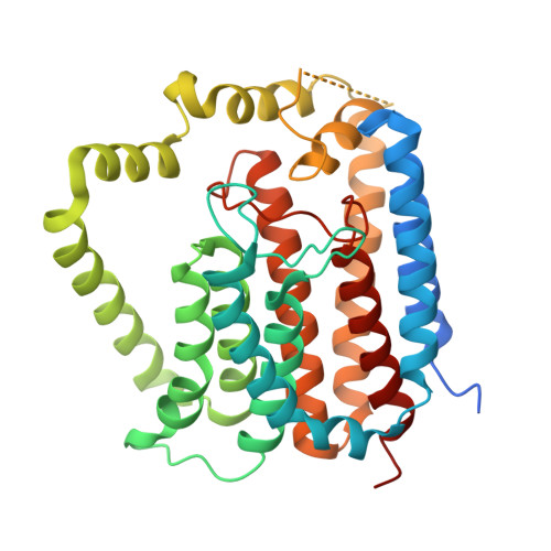
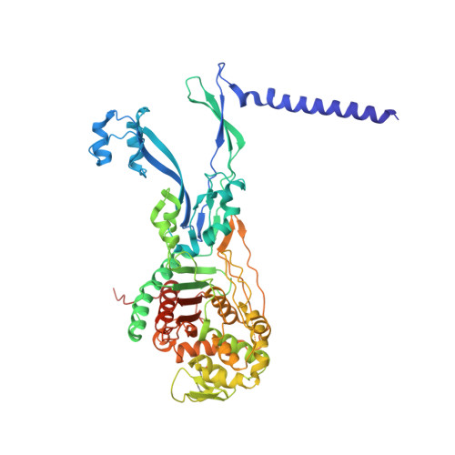
Structural coordination of polymerization and crosslinking by a SEDS-bPBP peptidoglycan synthase complex.

Sjodt, M.Rohs, P.D.A.Gilman, M.S.A.Erlandson, S.C.Zheng, S.Green, A.G.Brock, K.P.Taguchi, A.Kahne, D.Walker, S.Marks, D.S.Rudner, D.Z.Bernhardt, T.G.Kruse, A.C.

(2020) Nat Microbiol 5: 813-820

  • DOI: https://doi.org/10.1038/s41564-020-0687-z
  • Primary Citation of Related Structures:  
    6PL5, 6PL6

  • PubMed Abstract: 

    The shape, elongation, division and sporulation (SEDS) proteins are a highly conserved family of transmembrane glycosyltransferases that work in concert with class B penicillin-binding proteins (bPBPs) to build the bacterial peptidoglycan cell wall 1-6 . How these proteins coordinate polymerization of new glycan strands with their crosslinking to the existing peptidoglycan meshwork is unclear. Here, we report the crystal structure of the prototypical SEDS protein RodA from Thermus thermophilus in complex with its cognate bPBP at 3.3 Å resolution. The structure reveals a 1:1 stoichiometric complex with two extensive interaction interfaces between the proteins: one in the membrane plane and the other at the extracytoplasmic surface. When in complex with a bPBP, RodA shows an approximately 10 Å shift of transmembrane helix 7 that exposes a large membrane-accessible cavity. Negative-stain electron microscopy reveals that the complex can adopt a variety of different conformations. These data define the bPBP pedestal domain as the key allosteric activator of RodA both in vitro and in vivo, explaining how a SEDS-bPBP complex can coordinate its dual enzymatic activities of peptidoglycan polymerization and crosslinking to build the cell wall.


  • Organizational Affiliation

    Department of Biological Chemistry and Molecular Pharmacology, Blavatnik Institute, Harvard Medical School, Boston, MA, USA.


Macromolecules
Find similar proteins by:  (by identity cutoff)  |  3D Structure
Entity ID: 1
MoleculeChains Sequence LengthOrganismDetailsImage
Peptidoglycan glycosyltransferase RodA377Thermus thermophilus HB8Mutation(s): 0 
Gene Names: rodATTHA1241
EC: 2.4.1.129
Membrane Entity: Yes 
UniProt
Find proteins for Q5SIX3 (Thermus thermophilus (strain ATCC 27634 / DSM 579 / HB8))
Explore Q5SIX3 
Go to UniProtKB:  Q5SIX3
Entity Groups  
Sequence Clusters30% Identity50% Identity70% Identity90% Identity95% Identity100% Identity
UniProt GroupQ5SIX3
Sequence Annotations
Expand
  • Reference Sequence
Find similar proteins by:  (by identity cutoff)  |  3D Structure
Entity ID: 2
MoleculeChains Sequence LengthOrganismDetailsImage
Penicillin-binding protein 2/cell division protein FtsI598Thermus thermophilus HB8Mutation(s): 0 
Gene Names: TTHA1191
Membrane Entity: Yes 
UniProt
Find proteins for Q5SJ23 (Thermus thermophilus (strain ATCC 27634 / DSM 579 / HB8))
Explore Q5SJ23 
Go to UniProtKB:  Q5SJ23
Entity Groups  
Sequence Clusters30% Identity50% Identity70% Identity90% Identity95% Identity100% Identity
UniProt GroupQ5SJ23
Sequence Annotations
Expand
  • Reference Sequence

Find similar proteins by:  Sequence   |   3D Structure  

Entity ID: 3
MoleculeChains Sequence LengthOrganismDetailsImage
Unknown peptideC [auth D]11Thermus thermophilus HB8Mutation(s): 0 
Sequence Annotations
Expand
  • Reference Sequence
Experimental Data & Validation

Experimental Data

  • Method: X-RAY DIFFRACTION
  • Resolution: 3.50 Å
  • R-Value Free: 0.305 
  • R-Value Work: 0.285 
  • R-Value Observed: 0.287 
  • Space Group: P 32 2 1
Unit Cell:
Length ( Å )Angle ( ˚ )
a = 119.57α = 90
b = 119.57β = 90
c = 267.3γ = 120
Software Package:
Software NamePurpose
PHENIXrefinement
XDSdata reduction
XSCALEdata scaling
PHASERphasing
PDB_EXTRACTdata extraction

Structure Validation

View Full Validation Report


Entry History & Funding Information

Deposition Data


Funding OrganizationLocationGrant Number
National Institutes of Health/National Institute Of Allergy and Infectious Diseases (NIH/NIAID)United StatesU19AI109764

Revision History  (Full details and data files)

  • Version 1.0: 2020-03-18
    Type: Initial release
  • Version 1.1: 2020-03-25
    Changes: Database references
  • Version 1.2: 2020-06-10
    Changes: Database references
  • Version 1.3: 2023-10-11
    Changes: Data collection, Database references, Refinement description