6OR2

MmpL3 is a lipid transporter that binds trehalose monomycolate and phosphatidylethanolamine


Experimental Data Snapshot

  • Method: X-RAY DIFFRACTION
  • Resolution: 2.59 Å
  • R-Value Free: 0.290 
  • R-Value Work: 0.249 
  • R-Value Observed: 0.251 

wwPDB Validation   3D Report Full Report


Ligand Structure Quality Assessment 


This is version 1.4 of the entry. See complete history


Literature

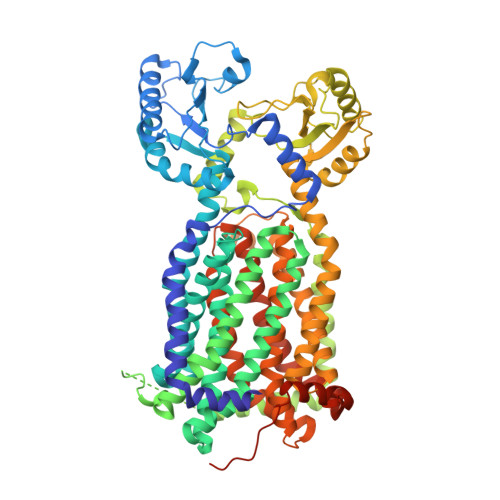
MmpL3 is a lipid transporter that binds trehalose monomycolate and phosphatidylethanolamine.

Su, C.C.Klenotic, P.A.Bolla, J.R.Purdy, G.E.Robinson, C.V.Yu, E.W.

(2019) Proc Natl Acad Sci U S A 116: 11241-11246

  • DOI: https://doi.org/10.1073/pnas.1901346116
  • Primary Citation of Related Structures:  
    6OR2

  • PubMed Abstract: 

    The cell envelope of Mycobacterium tuberculosis is notable for the abundance of mycolic acids (MAs), essential to mycobacterial viability, and of other species-specific lipids. The mycobacterial cell envelope is extremely hydrophobic, which contributes to virulence and antibiotic resistance. However, exactly how fatty acids and lipidic elements are transported across the cell envelope for cell-wall biosynthesis is unclear. Mycobacterial membrane protein Large 3 (MmpL3) is essential and required for transport of trehalose monomycolates (TMMs), precursors of MA-containing trehalose dimycolates (TDM) and mycolyl arabinogalactan peptidoglycan, but the exact function of MmpL3 remains elusive. Here, we report a crystal structure of Mycobacterium smegmatis MmpL3 at a resolution of 2.59 Å, revealing a monomeric molecule that is structurally distinct from all known bacterial membrane proteins. A previously unknown MmpL3 ligand, phosphatidylethanolamine (PE), was discovered inside this transporter. We also show, via native mass spectrometry, that MmpL3 specifically binds both TMM and PE, but not TDM, in the micromolar range. These observations provide insight into the function of MmpL3 and suggest a possible role for this protein in shuttling a variety of lipids to strengthen the mycobacterial cell wall.


  • Organizational Affiliation

    Department of Pharmacology, Case Western Reserve University School of Medicine, Cleveland, OH 44106.


Macromolecules
Find similar proteins by:  (by identity cutoff)  |  3D Structure
Entity ID: 1
MoleculeChains Sequence LengthOrganismDetailsImage
Membrane protein, MmpL family protein779Mycolicibacterium smegmatis MC2 155Mutation(s): 0 
Gene Names: MSMEG_0250
Membrane Entity: Yes 
UniProt
Find proteins for A0QP27 (Mycolicibacterium smegmatis (strain ATCC 700084 / mc(2)155))
Explore A0QP27 
Go to UniProtKB:  A0QP27
Entity Groups  
Sequence Clusters30% Identity50% Identity70% Identity90% Identity95% Identity100% Identity
UniProt GroupA0QP27
Sequence Annotations
Expand
  • Reference Sequence
Small Molecules
Ligands 2 Unique
IDChains Name / Formula / InChI Key2D Diagram3D Interactions
L9Q
Query on L9Q

Download Ideal Coordinates CCD File 
B [auth A](1S)-2-{[(S)-(2-aminoethoxy)(hydroxy)phosphoryl]oxy}-1-[(octadecanoyloxy)methyl]ethyl (9Z)-octadec-9-enoate
C41 H80 N O8 P
JQKOHRZNEOQNJE-VYCVAHKRSA-N
LMT
Query on LMT

Download Ideal Coordinates CCD File 
C [auth A]DODECYL-BETA-D-MALTOSIDE
C24 H46 O11
NLEBIOOXCVAHBD-QKMCSOCLSA-N
Experimental Data & Validation

Experimental Data

  • Method: X-RAY DIFFRACTION
  • Resolution: 2.59 Å
  • R-Value Free: 0.290 
  • R-Value Work: 0.249 
  • R-Value Observed: 0.251 
  • Space Group: P 21 21 21
Unit Cell:
Length ( Å )Angle ( ˚ )
a = 80.814α = 90
b = 129.457β = 90
c = 154.591γ = 90
Software Package:
Software NamePurpose
PHENIXrefinement
SCALEPACKdata scaling
PDB_EXTRACTdata extraction
DENZOdata reduction
PHENIXphasing

Structure Validation

View Full Validation Report


Ligand Structure Quality Assessment 


Entry History & Funding Information

Deposition Data

  • Released Date: 2019-05-29 
  • Deposition Author(s): Su, C.-C.
  • This entry supersedes: 6N3T

Funding OrganizationLocationGrant Number
National Institutes of Health/National Institute of General Medical Sciences (NIH/NIGMS)United States--

Revision History  (Full details and data files)

  • Version 1.0: 2019-05-29
    Type: Initial release
  • Version 1.1: 2019-06-05
    Changes: Data collection, Database references
  • Version 1.2: 2019-06-12
    Changes: Data collection, Database references
  • Version 1.3: 2020-01-01
    Changes: Author supporting evidence
  • Version 1.4: 2024-03-13
    Changes: Data collection, Database references