6O3P

Crystal structure of the catalytic domain of mouse Nudt12 in complex with AMP and 3 Mg2+ ions

  • Classification: HYDROLASE
  • Organism(s): Mus musculus
  • Expression System: Escherichia coli
  • Mutation(s): Yes 

  • Deposited: 2019-02-27 Released: 2019-05-29 
  • Deposition Author(s): Tong, L., Wu, Y.
  • Funding Organization(s): National Institutes of Health/National Human Genome Research Institute (NIH/NHGRI)

Experimental Data Snapshot

  • Method: X-RAY DIFFRACTION
  • Resolution: 1.60 Å
  • R-Value Free: 0.219 
  • R-Value Work: 0.180 
  • R-Value Observed: 0.181 

wwPDB Validation   3D Report Full Report


Ligand Structure Quality Assessment 


This is version 1.2 of the entry. See complete history


Literature

Structural and mechanistic basis of mammalian Nudt12 RNA deNADding.

Grudzien-Nogalska, E.Wu, Y.Jiao, X.Cui, H.Mateyak, M.K.Hart, R.P.Tong, L.Kiledjian, M.

(2019) Nat Chem Biol 15: 575-582

  • DOI: https://doi.org/10.1038/s41589-019-0293-7
  • Primary Citation of Related Structures:  
    6O3P

  • PubMed Abstract: 

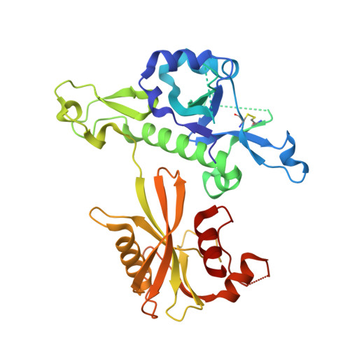
    We recently demonstrated that mammalian cells harbor nicotinamide adenine dinucleotide (NAD)-capped messenger RNAs that are hydrolyzed by the DXO deNADding enzyme. Here, we report that the Nudix protein Nudt12 is a second mammalian deNADding enzyme structurally and mechanistically distinct from DXO and targeting different RNAs. The crystal structure of mouse Nudt12 in complex with the deNADding product AMP and three Mg 2+ ions at 1.6 Å resolution provides insights into the molecular basis of the deNADding activity in the NAD pyrophosphate. Disruption of the Nudt12 gene stabilizes transfected NAD-capped RNA in cells, and its endogenous NAD-capped mRNA targets are enriched in those encoding proteins involved in cellular energetics. Furthermore, exposure of cells to nutrient or environmental stress manifests changes in NAD-capped RNA levels that are selectively responsive to Nudt12 or DXO, respectively, indicating an association of deNADding to cellular metabolism.


  • Organizational Affiliation

    Department Cell Biology and Neuroscience, Rutgers University, Piscataway, NJ, USA.


Macromolecules
Find similar proteins by:  (by identity cutoff)  |  3D Structure
Entity ID: 1
MoleculeChains Sequence LengthOrganismDetailsImage
Peroxisomal NADH pyrophosphatase NUDT12
A, B
339Mus musculusMutation(s): 3 
Gene Names: Nudt12
EC: 3.6.1.22
UniProt
Find proteins for Q9DCN1 (Mus musculus)
Explore Q9DCN1 
Go to UniProtKB:  Q9DCN1
Entity Groups  
Sequence Clusters30% Identity50% Identity70% Identity90% Identity95% Identity100% Identity
UniProt GroupQ9DCN1
Sequence Annotations
Expand
  • Reference Sequence
Small Molecules
Ligands 3 Unique
IDChains Name / Formula / InChI Key2D Diagram3D Interactions
AMP
Query on AMP

Download Ideal Coordinates CCD File 
C [auth A],
I [auth B]
ADENOSINE MONOPHOSPHATE
C10 H14 N5 O7 P
UDMBCSSLTHHNCD-KQYNXXCUSA-N
ZN
Query on ZN

Download Ideal Coordinates CCD File 
G [auth A],
H [auth B]
ZINC ION
Zn
PTFCDOFLOPIGGS-UHFFFAOYSA-N
MG
Query on MG

Download Ideal Coordinates CCD File 
D [auth A]
E [auth A]
F [auth A]
J [auth B]
K [auth B]
D [auth A],
E [auth A],
F [auth A],
J [auth B],
K [auth B],
L [auth B]
MAGNESIUM ION
Mg
JLVVSXFLKOJNIY-UHFFFAOYSA-N
Experimental Data & Validation

Experimental Data

  • Method: X-RAY DIFFRACTION
  • Resolution: 1.60 Å
  • R-Value Free: 0.219 
  • R-Value Work: 0.180 
  • R-Value Observed: 0.181 
  • Space Group: P 1
Unit Cell:
Length ( Å )Angle ( ˚ )
a = 56.207α = 102.4
b = 58.55β = 115.19
c = 61.707γ = 104.66
Software Package:
Software NamePurpose
PHENIXrefinement
XDSdata reduction
XDSdata scaling
PHENIXphasing

Structure Validation

View Full Validation Report


Ligand Structure Quality Assessment 


Entry History & Funding Information

Deposition Data


Funding OrganizationLocationGrant Number
National Institutes of Health/National Human Genome Research Institute (NIH/NHGRI)United States--

Revision History  (Full details and data files)

  • Version 1.0: 2019-05-29
    Type: Initial release
  • Version 1.1: 2019-12-18
    Changes: Author supporting evidence
  • Version 1.2: 2023-10-11
    Changes: Data collection, Database references, Derived calculations, Refinement description