6O3E

mouse aE-catenin 82-883

  • Classification: CELL ADHESION
  • Organism(s): Mus musculus
  • Expression System: Escherichia coli
  • Mutation(s): Yes 

  • Deposited: 2019-02-26 Released: 2019-11-13 
  • Deposition Author(s): Pokutta, S., Weis, W.I.
  • Funding Organization(s): National Institutes of Health/National Institute of General Medical Sciences (NIH/NIGMS)

Experimental Data Snapshot

  • Method: X-RAY DIFFRACTION
  • Resolution: 4.00 Å
  • R-Value Free: 0.243 
  • R-Value Work: 0.213 
  • R-Value Observed: 0.215 

wwPDB Validation   3D Report Full Report


This is version 1.2 of the entry. See complete history


Literature

Binding partner- and force-promoted changes in alpha E-catenin conformation probed by native cysteine labeling.

Terekhova, K.Pokutta, S.Kee, Y.S.Li, J.Tajkhorshid, E.Fuller, G.Dunn, A.R.Weis, W.I.

(2019) Sci Rep 9: 15375-15375

  • DOI: https://doi.org/10.1038/s41598-019-51816-3
  • Primary Citation of Related Structures:  
    6O3E

  • PubMed Abstract: 

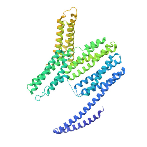
    Adherens Junctions (AJs) are cell-cell adhesion complexes that sense and propagate mechanical forces by coupling cadherins to the actin cytoskeleton via β-catenin and the F-actin binding protein αE-catenin. When subjected to mechanical force, the cadherin•catenin complex can tightly link to F-actin through αE-catenin, and also recruits the F-actin-binding protein vinculin. In this study, labeling of native cysteines combined with mass spectrometry revealed conformational changes in αE-catenin upon binding to the E-cadherin•β-catenin complex, vinculin and F-actin. A method to apply physiologically meaningful forces in solution revealed force-induced conformational changes in αE-catenin when bound to F-actin. Comparisons of wild-type αE-catenin and a mutant with enhanced vinculin affinity using cysteine labeling and isothermal titration calorimetry provide evidence for allosteric coupling of the N-terminal β-catenin-binding and the middle (M) vinculin-binding domain of αE-catenin. Cysteine labeling also revealed possible crosstalk between the actin-binding domain and the rest of the protein. The data provide insight into how binding partners and mechanical stress can regulate the conformation of full-length αE-catenin, and identify the M domain as a key transmitter of conformational changes.


  • Organizational Affiliation

    Departments of Structural Biology and Molecular & Cellular Physiology, Stanford University School of Medicine, Stanford, CA, 94305, USA.


Macromolecules
Find similar proteins by:  (by identity cutoff)  |  3D Structure
Entity ID: 1
MoleculeChains Sequence LengthOrganismDetailsImage
Catenin alpha-1
A, B
806Mus musculusMutation(s): 1 
Gene Names: Ctnna1Catna1
UniProt
Find proteins for P26231 (Mus musculus)
Explore P26231 
Go to UniProtKB:  P26231
Entity Groups  
Sequence Clusters30% Identity50% Identity70% Identity90% Identity95% Identity100% Identity
UniProt GroupP26231
Sequence Annotations
Expand
  • Reference Sequence
Experimental Data & Validation

Experimental Data

  • Method: X-RAY DIFFRACTION
  • Resolution: 4.00 Å
  • R-Value Free: 0.243 
  • R-Value Work: 0.213 
  • R-Value Observed: 0.215 
  • Space Group: P 32
Unit Cell:
Length ( Å )Angle ( ˚ )
a = 145.262α = 90
b = 145.262β = 90
c = 136.269γ = 120
Software Package:
Software NamePurpose
XDSdata reduction
Aimlessdata scaling
PHENIXrefinement
PDB_EXTRACTdata extraction
PHASERphasing

Structure Validation

View Full Validation Report


Entry History & Funding Information

Deposition Data


Funding OrganizationLocationGrant Number
National Institutes of Health/National Institute of General Medical Sciences (NIH/NIGMS)United StatesGM114462

Revision History  (Full details and data files)

  • Version 1.0: 2019-11-13
    Type: Initial release
  • Version 1.1: 2020-01-01
    Changes: Author supporting evidence
  • Version 1.2: 2023-10-11
    Changes: Data collection, Database references, Refinement description