


Structure of pCW3 conjugation coupling protein TcpA
Traore, D.A.K., Whisstock, J.C.To be published.
Experimental Data Snapshot
Entity ID: 1 | |||||
|---|---|---|---|---|---|
| Molecule | Chains | Sequence Length | Organism | Details | Image |
| DNA translocase coupling protein | 358 | Clostridium perfringens | Mutation(s): 0 Gene Names: tcpA, pCW3_0030 | 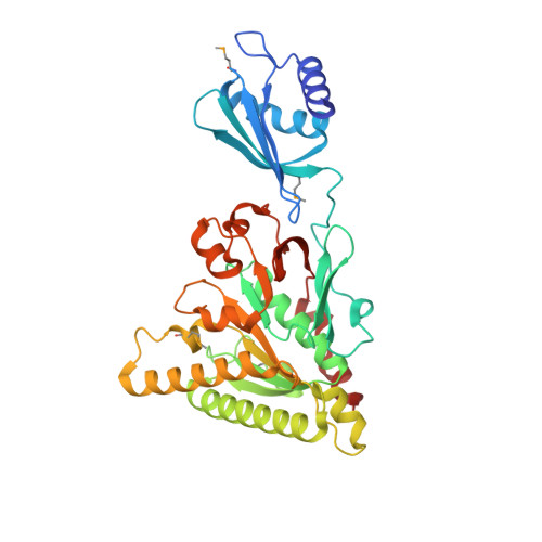 | |
UniProt | |||||
Find proteins for Q1PLI0 (Clostridium perfringens) Explore Q1PLI0 Go to UniProtKB: Q1PLI0 | |||||
Entity Groups | |||||
| Sequence Clusters | 30% Identity50% Identity70% Identity90% Identity95% Identity100% Identity | ||||
| UniProt Group | Q1PLI0 | ||||
Sequence AnnotationsExpand | |||||
| |||||
| Ligands 1 Unique | |||||
|---|---|---|---|---|---|
| ID | Chains | Name / Formula / InChI Key | 2D Diagram | 3D Interactions | |
| BGC Query on BGC | C [auth A], D [auth A], E [auth A], F [auth B], G [auth B] | beta-D-glucopyranose C6 H12 O6 WQZGKKKJIJFFOK-VFUOTHLCSA-N |  | ||
| Modified Residues 1 Unique | |||||
|---|---|---|---|---|---|
| ID | Chains | Type | Formula | 2D Diagram | Parent |
| MSE Query on MSE | A, B | L-PEPTIDE LINKING | C5 H11 N O2 Se |  | MET |
| Length ( Å ) | Angle ( ˚ ) |
|---|---|
| a = 53.17 | α = 90 |
| b = 101.83 | β = 90 |
| c = 140.04 | γ = 90 |
| Software Name | Purpose |
|---|---|
| BUSTER | refinement |
| Aimless | data scaling |
| SHELX | phasing |
| PDB_EXTRACT | data extraction |
| XDS | data reduction |
| Funding Organization | Location | Grant Number |
|---|---|---|
| National Health and Medical Research Council (NHMRC, Australia) | Australia | GNT1104502 |
RCSB PDB (citation) is hosted by
RCSB PDB is a member of the

