


Crystal structure of a full length elongation factor G (EF-G) from Rickettsia prowazekii
Edwards, T.E., Dranow, D.M., Lorimer, D.D., Horanyi, P.S., Seattle Structural Genomics Center for Infectious Disease (SSGCID)To be published.
Experimental Data Snapshot
wwPDB Validation 3D Report Full Report
Currently 6NOT does not have a validation slider image.
Entity ID: 1 | |||||
|---|---|---|---|---|---|
| Molecule | Chains | Sequence Length | Organism | Details | Image |
| Elongation factor G | 707 | Rickettsia prowazekii str. Madrid E | Mutation(s): 0 Gene Names: fusA, RP132 | 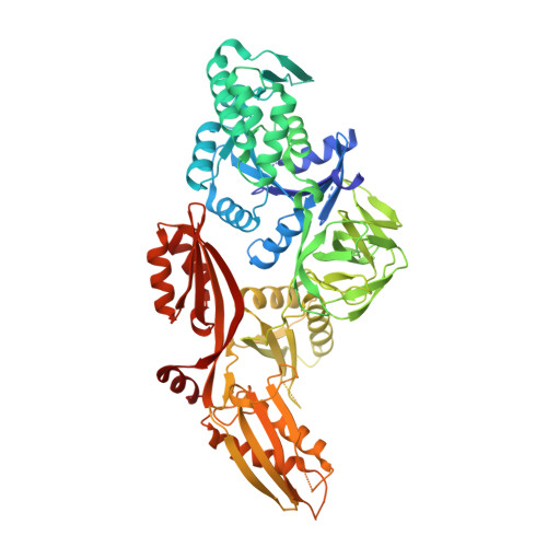 | |
UniProt | |||||
Find proteins for P41084 (Rickettsia prowazekii (strain Madrid E)) Explore P41084 Go to UniProtKB: P41084 | |||||
Entity Groups | |||||
| Sequence Clusters | 30% Identity50% Identity70% Identity90% Identity95% Identity100% Identity | ||||
| UniProt Group | P41084 | ||||
Sequence AnnotationsExpand | |||||
| |||||
| Length ( Å ) | Angle ( ˚ ) |
|---|---|
| a = 57.67 | α = 103.85 |
| b = 79.56 | β = 95.32 |
| c = 95.47 | γ = 92.17 |
| Software Name | Purpose |
|---|---|
| XDS | data reduction |
| XSCALE | data scaling |
| MOLREP | phasing |
| PHENIX | refinement |
| PDB_EXTRACT | data extraction |
Currently 6NOT does not have a validation slider image.
RCSB PDB (citation) is hosted by
RCSB PDB is a member of the

