6NHX

mycobacterial DNA ligase D complexed with ATP and MES


Experimental Data Snapshot

  • Method: X-RAY DIFFRACTION
  • Resolution: 1.40 Å
  • R-Value Free: 0.198 
  • R-Value Work: 0.188 
  • R-Value Observed: 0.188 

wwPDB Validation   3D Report Full Report


Ligand Structure Quality Assessment 


This is version 1.4 of the entry. See complete history


Literature

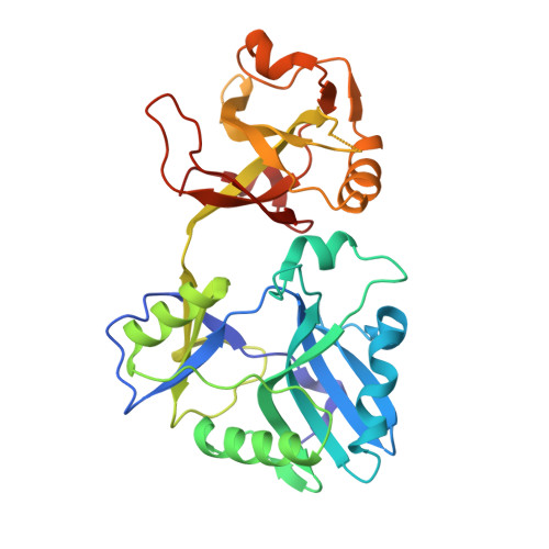
Structures of ATP-bound DNA ligase D in a closed domain conformation reveal a network of amino acid and metal contacts to the ATP phosphates.

Unciuleac, M.C.Goldgur, Y.Shuman, S.

(2019) J Biol Chem 294: 5094-5104

  • DOI: https://doi.org/10.1074/jbc.RA119.007445
  • Primary Citation of Related Structures:  
    6NHX, 6NHZ

  • PubMed Abstract: 

    DNA ligases are the sine qua non of genome integrity and essential for DNA replication and repair in all organisms. DNA ligases join 3'-OH and 5'-PO 4 ends via a series of three nucleotidyl transfer steps. In step 1, ligase reacts with ATP or NAD + to form a covalent ligase-(lysyl-Nζ)-AMP intermediate and release pyrophosphate (PP i ) or nicotinamide mononucleotide. In step 2, AMP is transferred from ligase-adenylate to the 5'-PO 4 DNA end to form a DNA-adenylate intermediate (AppDNA). In step 3, ligase catalyzes attack by a DNA 3'-OH on the DNA-adenylate to seal the two ends via a phosphodiester bond and release AMP. Eukaryal, archaeal, and many bacterial and viral DNA ligases are ATP-dependent. The catalytic core of ATP-dependent DNA ligases consists of an N-terminal nucleotidyltransferase domain fused to a C-terminal OB domain. Here we report crystal structures at 1.4-1.8 Å resolution of Mycobacterium tuberculosis LigD, an ATP-dependent DNA ligase dedicated to nonhomologous end joining, in complexes with ATP that highlight large movements of the OB domain (∼50 Å), from a closed conformation in the ATP complex to an open conformation in the covalent ligase-AMP intermediate. The LigD·ATP structures revealed a network of amino acid contacts to the ATP phosphates that stabilize the transition state and orient the PP i leaving group. A complex with ATP and magnesium suggested a two-metal mechanism of lysine adenylylation driven by a catalytic Mg 2+ that engages the ATP α phosphate and a second metal that bridges the ATP β and γ phosphates.


  • Organizational Affiliation

    From the Molecular Biology and.


Macromolecules
Find similar proteins by:  (by identity cutoff)  |  3D Structure
Entity ID: 1
MoleculeChains Sequence LengthOrganismDetailsImage
ATP-dependent DNA ligase307Mycobacterium tuberculosisMutation(s): 1 
Gene Names: ERS027656_00724
EC: 6.5.1.1
Entity Groups  
Sequence Clusters30% Identity50% Identity70% Identity90% Identity95% Identity100% Identity
Sequence Annotations
Expand
  • Reference Sequence
Small Molecules
Ligands 2 Unique
IDChains Name / Formula / InChI Key2D Diagram3D Interactions
ATP (Subject of Investigation/LOI)
Query on ATP

Download Ideal Coordinates CCD File 
B [auth A]ADENOSINE-5'-TRIPHOSPHATE
C10 H16 N5 O13 P3
ZKHQWZAMYRWXGA-KQYNXXCUSA-N
MES
Query on MES

Download Ideal Coordinates CCD File 
C [auth A]2-(N-MORPHOLINO)-ETHANESULFONIC ACID
C6 H13 N O4 S
SXGZJKUKBWWHRA-UHFFFAOYSA-N
Experimental Data & Validation

Experimental Data

  • Method: X-RAY DIFFRACTION
  • Resolution: 1.40 Å
  • R-Value Free: 0.198 
  • R-Value Work: 0.188 
  • R-Value Observed: 0.188 
  • Space Group: P 1 21 1
Unit Cell:
Length ( Å )Angle ( ˚ )
a = 41.089α = 90
b = 49.687β = 103.18
c = 73.522γ = 90
Software Package:
Software NamePurpose
PHENIXrefinement
HKL-2000data reduction
HKL-2000data scaling
MOLREPphasing

Structure Validation

View Full Validation Report


Ligand Structure Quality Assessment 


Entry History & Funding Information

Deposition Data


Funding OrganizationLocationGrant Number
National Institutes of Health/National Institute of General Medical Sciences (NIH/NIGMS)United StatesR35-GM126945
National Institutes of Health/National Cancer Institute (NIH/NCI)United StatesP30-CA008748

Revision History  (Full details and data files)

  • Version 1.0: 2019-02-13
    Type: Initial release
  • Version 1.1: 2019-02-20
    Changes: Data collection, Database references
  • Version 1.2: 2019-04-10
    Changes: Data collection, Database references
  • Version 1.3: 2019-12-04
    Changes: Author supporting evidence
  • Version 1.4: 2024-03-13
    Changes: Data collection, Database references