6MPV

Cryo-electron microscopy structure of Plasmodium falciparum Rh5/CyRPA/Ripr invasion complex


Experimental Data Snapshot

  • Method: ELECTRON MICROSCOPY
  • Resolution: 7.17 Å
  • Aggregation State: PARTICLE 
  • Reconstruction Method: SINGLE PARTICLE 

wwPDB Validation   3D Report Full Report


This is version 1.3 of the entry. See complete history


Literature

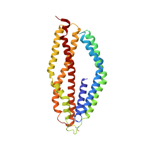
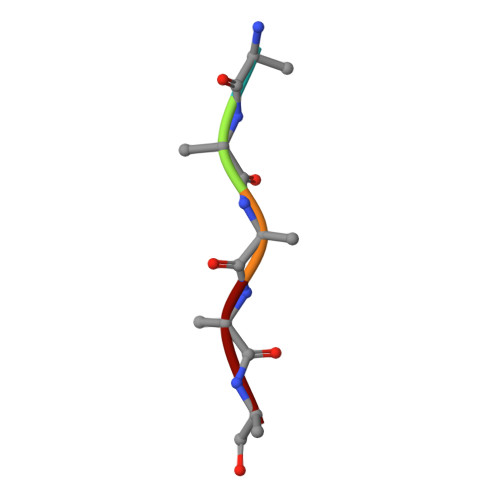
Structure of Plasmodium falciparum Rh5-CyRPA-Ripr invasion complex.

Wong, W.Huang, R.Menant, S.Hong, C.Sandow, J.J.Birkinshaw, R.W.Healer, J.Hodder, A.N.Kanjee, U.Tonkin, C.J.Heckmann, D.Soroka, V.Sogaard, T.M.M.Jorgensen, T.Duraisingh, M.T.Czabotar, P.E.de Jongh, W.A.Tham, W.H.Webb, A.I.Yu, Z.Cowman, A.F.

(2019) Nature 565: 118-121

  • DOI: https://doi.org/10.1038/s41586-018-0779-6
  • Primary Citation of Related Structures:  
    6MPV

  • PubMed Abstract: 

    Plasmodium falciparum causes the severe form of malaria that has high levels of mortality in humans. Blood-stage merozoites of P. falciparum invade erythrocytes, and this requires interactions between multiple ligands from the parasite and receptors in hosts. These interactions include the binding of the Rh5-CyRPA-Ripr complex with the erythrocyte receptor basigin 1,2 , which is an essential step for entry into human erythrocytes. Here we show that the Rh5-CyRPA-Ripr complex binds the erythrocyte cell line JK-1 significantly better than does Rh5 alone, and that this binding occurs through the insertion of Rh5 and Ripr into host membranes as a complex with high molecular weight. We report a cryo-electron microscopy structure of the Rh5-CyRPA-Ripr complex at subnanometre resolution, which reveals the organization of this essential invasion complex and the mode of interactions between members of the complex, and shows that CyRPA is a critical mediator of complex assembly. Our structure identifies blades 4-6 of the β-propeller of CyRPA as contact sites for Rh5 and Ripr. The limited contacts between Rh5-CyRPA and CyRPA-Ripr are consistent with the dissociation of Rh5 and Ripr from CyRPA for membrane insertion. A comparision of the crystal structure of Rh5-basigin with the cryo-electron microscopy structure of Rh5-CyRPA-Ripr suggests that Rh5 and Ripr are positioned parallel to the erythrocyte membrane before membrane insertion. This provides information on the function of this complex, and thereby provides insights into invasion by P. falciparum.


  • Organizational Affiliation

    Walter and Eliza Hall Institute of Medical Research, Parkville, Victoria, Australia.


Macromolecules
Find similar proteins by:  (by identity cutoff)  |  3D Structure
Entity ID: 1
MoleculeChains Sequence LengthOrganismDetailsImage
Cysteine-rich protective antigen332Plasmodium falciparum 3D7Mutation(s): 0 
Gene Names: PF3D7_0423800
UniProt
Find proteins for Q8IFM8 (Plasmodium falciparum (isolate 3D7))
Explore Q8IFM8 
Go to UniProtKB:  Q8IFM8
Entity Groups  
Sequence Clusters30% Identity50% Identity70% Identity90% Identity95% Identity100% Identity
UniProt GroupQ8IFM8
Sequence Annotations
Expand
  • Reference Sequence
Find similar proteins by:  (by identity cutoff)  |  3D Structure
Entity ID: 2
MoleculeChains Sequence LengthOrganismDetailsImage
Reticulocyte binding protein 5330Plasmodium falciparumMutation(s): 0 
UniProt
Find proteins for B2L3N7 (Plasmodium falciparum)
Explore B2L3N7 
Go to UniProtKB:  B2L3N7
Entity Groups  
Sequence Clusters30% Identity50% Identity70% Identity90% Identity95% Identity100% Identity
UniProt GroupB2L3N7
Sequence Annotations
Expand
  • Reference Sequence

Find similar proteins by:  Sequence   |   3D Structure  

Entity ID: 3
MoleculeChains Sequence LengthOrganismDetailsImage
PfRipr16Plasmodium falciparum 3D7Mutation(s): 0 
Sequence Annotations
Expand
  • Reference Sequence

Find similar proteins by:  Sequence   |   3D Structure  

Entity ID: 4
MoleculeChains Sequence LengthOrganismDetailsImage
PfRipr5Plasmodium falciparum 3D7Mutation(s): 0 
Sequence Annotations
Expand
  • Reference Sequence
Experimental Data & Validation

Experimental Data

  • Method: ELECTRON MICROSCOPY
  • Resolution: 7.17 Å
  • Aggregation State: PARTICLE 
  • Reconstruction Method: SINGLE PARTICLE 
EM Software:
TaskSoftware PackageVersion
MODEL REFINEMENTPHENIX

Structure Validation

View Full Validation Report


Entry History & Funding Information

Deposition Data


Funding OrganizationLocationGrant Number
National Health and Medical Research Council (NHMRC, Australia)AustraliaGNT637406

Revision History  (Full details and data files)

  • Version 1.0: 2018-12-12
    Type: Initial release
  • Version 1.1: 2018-12-26
    Changes: Data collection, Database references
  • Version 1.2: 2019-01-16
    Changes: Data collection, Database references
  • Version 1.3: 2019-12-18
    Changes: Author supporting evidence, Other