Identification of structural transitions in bacterial fatty acid binding proteins that permit ligand entry and exit at membranes.
Gullett, J.M., Cuypers, M.G., Grace, C.R., Pant, S., Subramanian, C., Tajkhorshid, E., Rock, C.O., White, S.W.(2022) J Biol Chem 298: 101676-101676
- PubMed: 35122790
- DOI: https://doi.org/10.1016/j.jbc.2022.101676
- Primary Citation of Related Structures:
6MH9, 6NM1, 7SCL, 7SG3 - PubMed Abstract:
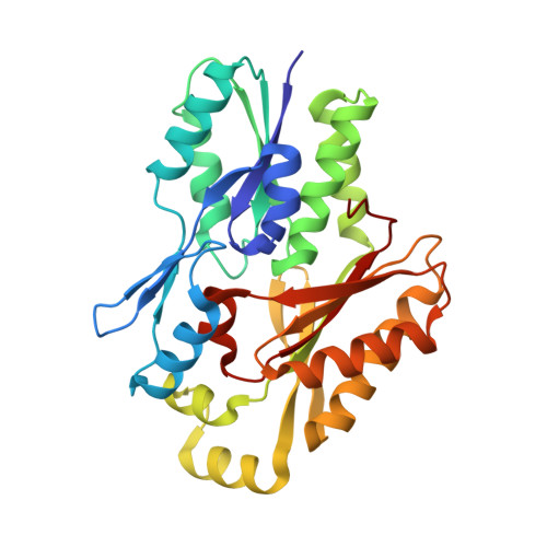
Fatty acid (FA) transfer proteins extract FA from membranes and sequester them to facilitate their movement through the cytosol. Detailed structural information is available for these soluble protein-FA complexes, but the structure of the protein conformation responsible for FA exchange at the membrane is unknown. Staphylococcus aureus FakB1 is a prototypical bacterial FA transfer protein that binds palmitate within a narrow, buried tunnel. Here, we define the conformational change from a "closed" FakB1 state to an "open" state that associates with the membrane and provides a path for entry and egress of the FA. Using NMR spectroscopy, we identified a conformationally flexible dynamic region in FakB1, and X-ray crystallography of FakB1 mutants captured the conformation of the open state. In addition, molecular dynamics simulations show that the new amphipathic α-helix formed in the open state inserts below the phosphate plane of the bilayer to create a diffusion channel for the hydrophobic FA tail to access the hydrocarbon core and place the carboxyl group at the phosphate layer. The membrane binding and catalytic properties of site-directed mutants were consistent with the proposed membrane docked structure predicted by our molecular dynamics simulations. Finally, the structure of the bilayer-associated conformation of FakB1 has local similarities with mammalian FA binding proteins and provides a conceptual framework for how these proteins interact with the membrane to create a diffusion channel from the FA location in the bilayer to the protein interior.
Organizational Affiliation:
Department of Infectious Diseases, St. Jude Children's Research Hospital, Memphis, Tennessee, USA.