6MCJ

Structure of Helical Carotenoid Protein 2 from Fremyella diplosiphon


Experimental Data Snapshot

  • Method: X-RAY DIFFRACTION
  • Resolution: 1.71 Å
  • R-Value Free: 0.199 
  • R-Value Work: 0.167 
  • R-Value Observed: 0.169 

wwPDB Validation   3D Report Full Report

Currently 6MCJ does not have a validation slider image.


This is version 1.2 of the entry. See complete history


Literature

Structural and spectroscopic characterization of HCP2.

Dominguez-Martin, M.A.Polivka, T.Sutter, M.Ferlez, B.Lechno-Yossef, S.Montgomery, B.L.Kerfeld, C.A.

(2019) Biochim Biophys Acta Bioenerg 1860: 414-424

  • DOI: https://doi.org/10.1016/j.bbabio.2019.03.004
  • Primary Citation of Related Structures:  
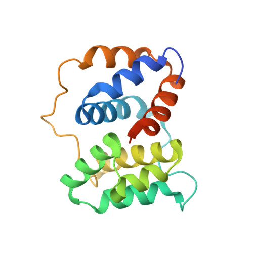
    6MCJ

  • PubMed Abstract: 

    The Helical Carotenoid Proteins (HCPs) are a large group of newly identified carotenoid-binding proteins found in ecophysiologically diverse cyanobacteria. They likely evolved before becoming the effector (quenching) domain of the modular Orange Carotenoid Protein (OCP). The number of discrete HCP families-at least nine-suggests they are involved in multiple distinct functions. Here we report the 1.7 Å crystal structure of HCP2, one of the most widespread HCPs found in nature, from the chromatically acclimating cyanobacterium Tolypothrix sp. PCC 7601. By purifying HCP2 from the native source we are able to identify its natively-bound carotenoid, which is exclusively canthaxanthin. In solution, HCP2 is a monomer with an absorbance maximum of 530 nm. However, the HCP2 crystals have a maximum absorbance at 548 nm, which is accounted by the stacking of the β1 rings of the carotenoid in the two molecules in the asymmetric unit. Our results demonstrate how HCPs provide a valuable system to study carotenoid-protein interactions and their spectroscopic implications, and contribute to efforts to understand the functional roles of this large, newly discovered family of pigment proteins, which to-date remain enigmatic.


  • Organizational Affiliation

    MSU-DOE Plant Research Laboratory, Michigan State University, East Lansing, MI 48824, USA.


Macromolecules
Find similar proteins by:  (by identity cutoff)  |  3D Structure
Entity ID: 1
MoleculeChains Sequence LengthOrganismDetailsImage
Orange carotenoid-binding protein
A, B
163Tolypothrix sp. PCC 7601Mutation(s): 0 
Gene Names: FDUTEX481_01662
UniProt
Find proteins for A0A0D6L0Q7 (Tolypothrix sp. PCC 7601)
Explore A0A0D6L0Q7 
Go to UniProtKB:  A0A0D6L0Q7
Entity Groups  
Sequence Clusters30% Identity50% Identity70% Identity90% Identity95% Identity100% Identity
UniProt GroupA0A0D6L0Q7
Sequence Annotations
Expand
  • Reference Sequence
Small Molecules
Ligands 3 Unique
IDChains Name / Formula / InChI Key2D Diagram3D Interactions
45D
Query on 45D

Download Ideal Coordinates CCD File 
C [auth A],
I [auth B]
beta,beta-carotene-4,4'-dione
C40 H52 O2
FDSDTBUPSURDBL-DKLMTRRASA-N
1PE
Query on 1PE

Download Ideal Coordinates CCD File 
H [auth A]PENTAETHYLENE GLYCOL
C10 H22 O6
JLFNLZLINWHATN-UHFFFAOYSA-N
IOD
Query on IOD

Download Ideal Coordinates CCD File 
D [auth A]
E [auth A]
F [auth A]
G [auth A]
J [auth B]
D [auth A],
E [auth A],
F [auth A],
G [auth A],
J [auth B],
K [auth B],
L [auth B],
M [auth B]
IODIDE ION
I
XMBWDFGMSWQBCA-UHFFFAOYSA-M
Experimental Data & Validation

Experimental Data

  • Method: X-RAY DIFFRACTION
  • Resolution: 1.71 Å
  • R-Value Free: 0.199 
  • R-Value Work: 0.167 
  • R-Value Observed: 0.169 
  • Space Group: P 1 21 1
Unit Cell:
Length ( Å )Angle ( ˚ )
a = 35.162α = 90
b = 71.635β = 92.32
c = 56.341γ = 90
Software Package:
Software NamePurpose
PHENIXrefinement
XDSdata reduction
SCALAdata scaling
PHASERphasing

Structure Validation

View Full Validation Report

Currently 6MCJ does not have a validation slider image.


Entry History 

Deposition Data

Revision History  (Full details and data files)

  • Version 1.0: 2019-03-27
    Type: Initial release
  • Version 1.1: 2019-11-13
    Changes: Database references
  • Version 1.2: 2023-10-11
    Changes: Data collection, Database references, Refinement description