6LK1

Ultrahigh resolution X-ray structure of Ferredoxin I from C. reinhardtii


Experimental Data Snapshot

  • Method: X-RAY DIFFRACTION
  • Resolution: 0.90 Å
  • R-Value Free: 0.171 
  • R-Value Work: 0.145 

wwPDB Validation   3D Report Full Report


This is version 1.2 of the entry. See complete history


Literature

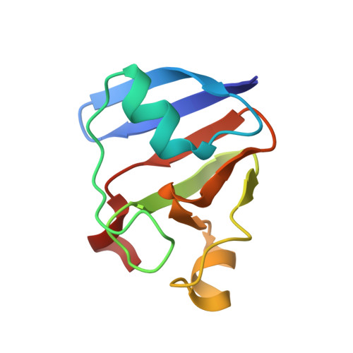
X-ray dose-dependent structural changes of the [2Fe-2S] ferredoxin from Chlamydomonas reinhardtii.

Ohnishi, Y.Muraki, N.Kiyota, D.Okumura, H.Baba, S.Kawano, Y.Kumasaka, T.Tanaka, H.Kurisu, G.

(2020) J Biochem 167: 549-555

  • DOI: https://doi.org/10.1093/jb/mvaa045
  • Primary Citation of Related Structures:  
    6KUM, 6KV0, 6LK1

  • PubMed Abstract: 

    Plant-type ferredoxin (Fd) is an electron transfer protein in chloroplast. Redox-dependent structural change of Fd controls its association with and dissociation from Fd-dependent enzymes. Among many X-ray structures of oxidized Fd have been reported so far, very likely a given number of them was partially reduced by strong X-ray. To understand the precise structural change between reduced and oxidized Fd, it is important to know whether the crystals of oxidized Fd may or may not be reduced during the X-ray experiment. We prepared the thin plate-shaped Fd crystals from Chlamydomonas reinhardtii and monitored its absorption spectra during experiment. Absorption spectra of oxidized Fd crystals were clearly changed to that of reduced form in an X-ray dose-dependent manner. In another independent experiment, the X-ray diffraction images obtained from different parts of one single crystal were sorted and merged to form two datasets with low and high X-ray doses. An Fo-Fo map calculated from the two datasets showed that X-ray reduction causes a small displacement of the iron atoms in the [2Fe-2S] cluster. Both our spectroscopic and crystallographic studies confirm X-ray dose-dependent reduction of Fd, and suggest a structural basis for its initial reduction step especially in the core of the cluster.


  • Organizational Affiliation

    Division of Protein Structural Biology, Institute for Protein Research, Osaka University, 3-2 Yamadaoka, Suita, Osaka 565-0871, Japan.


Macromolecules
Find similar proteins by:  (by identity cutoff)  |  3D Structure
Entity ID: 1
MoleculeChains Sequence LengthOrganismDetailsImage
Ferredoxin, chloroplastic
A, B
95Chlamydomonas reinhardtiiMutation(s): 0 
Gene Names: PETF
UniProt
Find proteins for P07839 (Chlamydomonas reinhardtii)
Explore P07839 
Go to UniProtKB:  P07839
Entity Groups  
Sequence Clusters30% Identity50% Identity70% Identity90% Identity95% Identity100% Identity
UniProt GroupP07839
Sequence Annotations
Expand
  • Reference Sequence
Small Molecules
Ligands 4 Unique
IDChains Name / Formula / InChI Key2D Diagram3D Interactions
FES (Subject of Investigation/LOI)
Query on FES

Download Ideal Coordinates CCD File 
C [auth A],
L [auth B]
FE2/S2 (INORGANIC) CLUSTER
Fe2 S2
NIXDOXVAJZFRNF-UHFFFAOYSA-N
BEN
Query on BEN

Download Ideal Coordinates CCD File 
D [auth A]
E [auth A]
F [auth A]
G [auth A]
H [auth A]
D [auth A],
E [auth A],
F [auth A],
G [auth A],
H [auth A],
I [auth A],
M [auth B],
N [auth B],
O [auth B],
P [auth B]
BENZAMIDINE
C7 H8 N2
PXXJHWLDUBFPOL-UHFFFAOYSA-N
SO4
Query on SO4

Download Ideal Coordinates CCD File 
K [auth A]SULFATE ION
O4 S
QAOWNCQODCNURD-UHFFFAOYSA-L
SCN
Query on SCN

Download Ideal Coordinates CCD File 
J [auth A]THIOCYANATE ION
C N S
ZMZDMBWJUHKJPS-UHFFFAOYSA-M
Experimental Data & Validation

Experimental Data

  • Method: X-RAY DIFFRACTION
  • Resolution: 0.90 Å
  • R-Value Free: 0.171 
  • R-Value Work: 0.145 
  • Space Group: P 1 21 1
Unit Cell:
Length ( Å )Angle ( ˚ )
a = 25.955α = 90
b = 60.09β = 102.4
c = 50.085γ = 90
Software Package:
Software NamePurpose
SHELXrefinement
PDB_EXTRACTdata extraction
MOSFLMdata reduction
Aimlessdata scaling
PHENIXphasing

Structure Validation

View Full Validation Report


Entry History & Funding Information

Deposition Data


Funding OrganizationLocationGrant Number
Ministry of Education, Culture, Sports, Science and Technology (Japan)Japan--

Revision History  (Full details and data files)

  • Version 1.0: 2020-05-27
    Type: Initial release
  • Version 1.1: 2020-06-10
    Changes: Database references
  • Version 1.2: 2023-11-22
    Changes: Data collection, Database references, Refinement description