6KUN

Crystal structure of dioxygenase for auxin oxidation (DAO) in rice


Experimental Data Snapshot

  • Method: X-RAY DIFFRACTION
  • Resolution: 2.00 Å
  • R-Value Free: 0.243 
  • R-Value Work: 0.188 
  • R-Value Observed: 0.191 

wwPDB Validation   3D Report Full Report


Ligand Structure Quality Assessment 


This is version 1.1 of the entry. See complete history


Literature

A common allosteric mechanism regulates homeostatic inactivation of auxin and gibberellin.

Takehara, S.Sakuraba, S.Mikami, B.Yoshida, H.Yoshimura, H.Itoh, A.Endo, M.Watanabe, N.Nagae, T.Matsuoka, M.Ueguchi-Tanaka, M.

(2020) Nat Commun 11: 2143-2143

  • DOI: https://doi.org/10.1038/s41467-020-16068-0
  • Primary Citation of Related Structures:  
    6KU3, 6KUN

  • PubMed Abstract: 

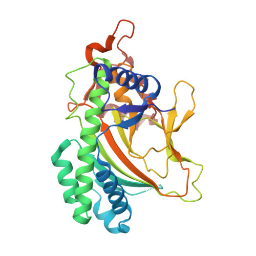
    Allosteric regulation is protein activation by effector binding at a site other than the active site. Here, we show via X-ray structural analysis of gibberellin 2-oxidase 3 (GA2ox3), and auxin dioxygenase (DAO), that such a mechanism maintains hormonal homeostasis in plants. Both enzymes form multimers by interacting via GA 4 and indole-3-acetic acid (IAA) at their binding interface. Via further functional analyses we reveal that multimerization of these enzymes gradually proceeds with increasing GA 4 and IAA concentrations; multimerized enzymes have higher specific activities than monomer forms, a system that should favour the maintenance of homeostasis for these phytohormones. Molecular dynamic analysis suggests a possible mechanism underlying increased GA2ox3 activity by multimerization-GA 4 in the interface of oligomerized GA2ox3s may be able to enter the active site with a low energy barrier. In summary, homeostatic systems for maintaining GA and IAA levels, based on a common allosteric mechanism, appear to have developed independently.


  • Organizational Affiliation

    Bioscience and Biotechnology Centre, Nagoya University, Nagoya, 464-8601, Japan.


Macromolecules
Find similar proteins by:  (by identity cutoff)  |  3D Structure
Entity ID: 1
MoleculeChains Sequence LengthOrganismDetailsImage
2-oxoglutarate-dependent dioxygenase DAO
A, B
300Oryza sativa Indica GroupMutation(s): 0 
Gene Names: DAOOsI_16300OSIGBa0106G07.8
EC: 1.14.11
UniProt
Find proteins for Q01IX6 (Oryza sativa subsp. indica)
Explore Q01IX6 
Go to UniProtKB:  Q01IX6
Entity Groups  
Sequence Clusters30% Identity50% Identity70% Identity90% Identity95% Identity100% Identity
UniProt GroupQ01IX6
Sequence Annotations
Expand
  • Reference Sequence
Small Molecules
Ligands 3 Unique
IDChains Name / Formula / InChI Key2D Diagram3D Interactions
IAC (Subject of Investigation/LOI)
Query on IAC

Download Ideal Coordinates CCD File 
D [auth A],
H [auth B]
1H-INDOL-3-YLACETIC ACID
C10 H9 N O2
SEOVTRFCIGRIMH-UHFFFAOYSA-N
AKG
Query on AKG

Download Ideal Coordinates CCD File 
C [auth A],
G [auth B]
2-OXOGLUTARIC ACID
C5 H6 O5
KPGXRSRHYNQIFN-UHFFFAOYSA-N
GOL
Query on GOL

Download Ideal Coordinates CCD File 
E [auth A],
F [auth A],
I [auth B],
J [auth B]
GLYCEROL
C3 H8 O3
PEDCQBHIVMGVHV-UHFFFAOYSA-N
Experimental Data & Validation

Experimental Data

  • Method: X-RAY DIFFRACTION
  • Resolution: 2.00 Å
  • R-Value Free: 0.243 
  • R-Value Work: 0.188 
  • R-Value Observed: 0.191 
  • Space Group: P 21 21 21
Unit Cell:
Length ( Å )Angle ( ˚ )
a = 75.247α = 90
b = 78.304β = 90
c = 94.003γ = 90
Software Package:
Software NamePurpose
PHENIXrefinement
Cootmodel building
HKL-2000data reduction
HKL-2000data scaling
MOLREPphasing

Structure Validation

View Full Validation Report


Ligand Structure Quality Assessment 


Entry History & Funding Information

Deposition Data


Funding OrganizationLocationGrant Number
Japan Society for the Promotion of Science (JSPS)Japan16H06468
Japan Society for the Promotion of Science (JSPS)Japan16H06464

Revision History  (Full details and data files)

  • Version 1.0: 2020-05-13
    Type: Initial release
  • Version 1.1: 2023-11-22
    Changes: Data collection, Database references, Refinement description