6KNK

Crystal structure of SbnH in complex with citryl-diaminoethane


Experimental Data Snapshot

  • Method: X-RAY DIFFRACTION
  • Resolution: 2.30 Å
  • R-Value Free: 0.245 
  • R-Value Work: 0.221 
  • R-Value Observed: 0.222 

wwPDB Validation   3D Report Full Report


Ligand Structure Quality Assessment 


This is version 1.2 of the entry. See complete history


Literature

Structural Insights into Substrate Recognition and Activity Regulation of the Key Decarboxylase SbnH in Staphyloferrin B Biosynthesis.

Tang, J.Ju, Y.Gu, Q.Xu, J.Zhou, H.

(2019) J Mol Biol 431: 4868-4881

  • DOI: https://doi.org/10.1016/j.jmb.2019.10.009
  • Primary Citation of Related Structures:  
    6KNH, 6KNI, 6KNK

  • PubMed Abstract: 

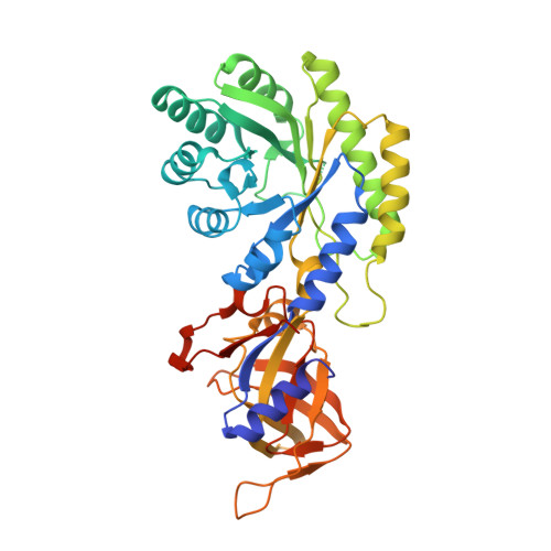
    Staphyloferrin B is a hydroxycarboxylate siderophore that is crucial for the invasion and virulence of Staphylococcus aureus in mammalian hosts where free iron ions are scarce. The assembly of staphyloferrin B involves four enzymatic steps, in which SbnH, a pyridoxal 5'-phosphate (PLP)-dependent decarboxylase, catalyzes the second step. Here, we report the X-ray crystal structures of S. aureus SbnH (SaSbnH) in complex with PLP, citrate, and the decarboxylation product citryl-diaminoethane (citryl-Dae). The overall structure of SaSbnH resembles those of the previously reported PLP-dependent amino acid decarboxylases, but the active site of SaSbnH showed unique structural features. Structural and mutagenesis analysis revealed that the citryl moiety of the substrate citryl-l-2,3-diaminopropionic acid (citryl-l-Dap) inserts into a narrow groove at the dimer interface of SaSbnH and forms hydrogen bonding interactions with both subunits. In the active site, a conserved lysine residue forms an aldimine linkage with the cofactor PLP, and a phenylalanine residue is essential for accommodating the l-configuration Dap of the substrate. Interestingly, the freestanding citrate molecule was found to bind to SaSbnH in a conformation inverse to that of the citryl group of citryl-Dae and efficiently inhibit SaSbnH. As an intermediate in the tricarboxylic acid (TCA) cycle, citrate is highly abundant in bacterial cells until iron depletion; thus, its inhibition of SaSbnH may serve as an iron-dependent regulatory mechanism in staphyloferrin B biosynthesis.


  • Organizational Affiliation

    Guangdong Provincial Key Laboratory of Chiral Molecule and Drug Discovery, School of Pharmaceutical Sciences, Sun Yat-sen University, Guangzhou 510006, China; Research Center for Drug Discovery, School of Pharmaceutical Sciences, Sun Yat-sen University, Guangzhou 510006, China.


Macromolecules
Find similar proteins by:  (by identity cutoff)  |  3D Structure
Entity ID: 1
MoleculeChains Sequence LengthOrganismDetailsImage
Probable diaminopimelate decarboxylase protein
A, B, C
406Staphylococcus aureus subsp. aureus Mu50Mutation(s): 1 
Gene Names: SAV0123
UniProt
Find proteins for A0A0H3JPF2 (Staphylococcus aureus (strain Mu50 / ATCC 700699))
Explore A0A0H3JPF2 
Go to UniProtKB:  A0A0H3JPF2
Entity Groups  
Sequence Clusters30% Identity50% Identity70% Identity90% Identity95% Identity100% Identity
UniProt GroupA0A0H3JPF2
Sequence Annotations
Expand
  • Reference Sequence
Small Molecules
Ligands 3 Unique
IDChains Name / Formula / InChI Key2D Diagram3D Interactions
DN9 (Subject of Investigation/LOI)
Query on DN9

Download Ideal Coordinates CCD File 
D [auth A](2~{S})-2-[2-[2-[[2-methyl-3-oxidanyl-5-(phosphonooxymethyl)pyridin-4-yl]methylamino]ethylamino]-2-oxidanylidene-ethyl]-2-oxidanyl-butanedioic acid
C16 H24 N3 O11 P
LHKUXYWSSPZJHQ-INIZCTEOSA-N
X3J (Subject of Investigation/LOI)
Query on X3J

Download Ideal Coordinates CCD File 
F [auth B](2S)-2-{2-[(2-AMINOETHYL)AMINO]-2-OXOETHYL}-2-HYDROXYBUTANEDIOIC ACID
C8 H14 N2 O6
KQWWDTIPDRWKGZ-QMMMGPOBSA-N
PO4
Query on PO4

Download Ideal Coordinates CCD File 
E [auth B],
G [auth C]
PHOSPHATE ION
O4 P
NBIIXXVUZAFLBC-UHFFFAOYSA-K
Experimental Data & Validation

Experimental Data

  • Method: X-RAY DIFFRACTION
  • Resolution: 2.30 Å
  • R-Value Free: 0.245 
  • R-Value Work: 0.221 
  • R-Value Observed: 0.222 
  • Space Group: C 1 2 1
Unit Cell:
Length ( Å )Angle ( ˚ )
a = 176.299α = 90
b = 80.325β = 102.07
c = 111.524γ = 90
Software Package:
Software NamePurpose
REFMACrefinement
XDSdata reduction
Aimlessdata scaling
MOLREPphasing

Structure Validation

View Full Validation Report


Ligand Structure Quality Assessment 


Entry History & Funding Information

Deposition Data


Funding OrganizationLocationGrant Number
Ministry of Science and Technology (China)China2017YFE0109900
National Natural Science Foundation of ChinaChinaNo. 81773636

Revision History  (Full details and data files)

  • Version 1.0: 2019-11-13
    Type: Initial release
  • Version 1.1: 2020-01-01
    Changes: Database references
  • Version 1.2: 2023-11-22
    Changes: Data collection, Database references, Refinement description