


Crystal structure of hydrogen peroxide bound bovine lactoperoxidase at 2.3 A resolution
Singh, P.K., Sirohi, H.V., Bhushan, A., Kaur, P., Sharma, S., Singh, T.P.To be published.
Experimental Data Snapshot
Entity ID: 1 | |||||
|---|---|---|---|---|---|
| Molecule | Chains | Sequence Length | Organism | Details | Image |
| Lactoperoxidase | 595 | Bos taurus | Mutation(s): 0 EC: 1.11.1.7 | 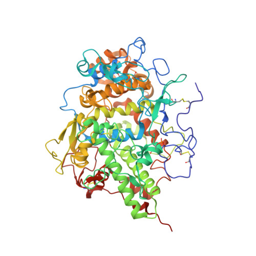 | |
UniProt | |||||
Find proteins for P80025 (Bos taurus) Explore P80025 Go to UniProtKB: P80025 | |||||
Entity Groups | |||||
| Sequence Clusters | 30% Identity50% Identity70% Identity90% Identity95% Identity100% Identity | ||||
| UniProt Group | P80025 | ||||
Sequence AnnotationsExpand | |||||
| |||||
| Ligands 5 Unique | |||||
|---|---|---|---|---|---|
| ID | Chains | Name / Formula / InChI Key | 2D Diagram | 3D Interactions | |
| HEM (Subject of Investigation/LOI) Query on HEM | B [auth A] | PROTOPORPHYRIN IX CONTAINING FE C34 H32 Fe N4 O4 KABFMIBPWCXCRK-RGGAHWMASA-L |  | ||
| NAG (Subject of Investigation/LOI) Query on NAG | D [auth A], E [auth A], F [auth A], G [auth A] | 2-acetamido-2-deoxy-beta-D-glucopyranose C8 H15 N O6 OVRNDRQMDRJTHS-FMDGEEDCSA-N |  | ||
| IOD Query on IOD | H [auth A] I [auth A] J [auth A] K [auth A] L [auth A] | IODIDE ION I XMBWDFGMSWQBCA-UHFFFAOYSA-M |  | ||
| CA Query on CA | C [auth A] | CALCIUM ION Ca BHPQYMZQTOCNFJ-UHFFFAOYSA-N |  | ||
| PEO Query on PEO | W [auth A], X [auth A] | HYDROGEN PEROXIDE H2 O2 MHAJPDPJQMAIIY-UHFFFAOYSA-N |  | ||
| Modified Residues 1 Unique | |||||
|---|---|---|---|---|---|
| ID | Chains | Type | Formula | 2D Diagram | Parent |
| SEP Query on SEP | A | L-PEPTIDE LINKING | C3 H8 N O6 P |  | SER |
| Length ( Å ) | Angle ( ˚ ) |
|---|---|
| a = 53.828 | α = 90 |
| b = 80.42 | β = 102.577 |
| c = 75.823 | γ = 90 |
| Software Name | Purpose |
|---|---|
| REFMAC | refinement |
| AUTOMAR | data reduction |
| Aimless | data scaling |
| MOLREP | phasing |
RCSB PDB (citation) is hosted by
RCSB PDB is a member of the

