6KLK

Crystal structure of the Pseudomonas aeruginosa dihydropyrimidinase complexed with 5-FU


Experimental Data Snapshot

  • Method: X-RAY DIFFRACTION
  • Resolution: 1.76 Å
  • R-Value Free: 0.212 
  • R-Value Work: 0.185 
  • R-Value Observed: 0.187 

wwPDB Validation   3D Report Full Report


Ligand Structure Quality Assessment 


This is version 1.1 of the entry. See complete history


Literature

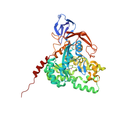
Crystal structure of dihydropyrimidinase in complex with anticancer drug 5-fluorouracil.

Huang, Y.H.Ning, Z.J.Huang, C.Y.

(2019) Biochem Biophys Res Commun 519: 160-165

  • DOI: https://doi.org/10.1016/j.bbrc.2019.08.153
  • Primary Citation of Related Structures:  
    6KLK

  • PubMed Abstract: 

    Dihydropyrimidinase (DHPase) catalyzes the reversible cyclization of dihydrouracil to N-carbamoyl-β-alanine in the second step of the pyrimidine degradation pathway. Whether 5-fluorouracil (5-FU), the best-known fluoropyrimidine that is used to target the enzyme thymidylate synthase for anticancer therapy, can bind to DHPase remains unknown. In this study, we found that 5-FU can form a stable complex with Pseudomonas aeruginosa DHPase (PaDHPase). The crystal structure of PaDHPase complexed with 5-FU was determined at 1.76 Å resolution (PDB entry 6KLK). Various interactions between 5-FU and PaDHPase were examined. Six residues, namely, His61, Tyr155, Asp316, Cys318, Ser289 and Asn337, of PaDHPase were involved in 5-FU binding. Except for Cys318, these residues are also known as the substrate-binding sites of DHPase. 5-FU interacts with the main chains of residues Ser289 (3.0 Å) and Asn337 (3.2 Å) and the side chains of residues Tyr155 (2.8 Å) and Cys318 (2.9 Å). Mutation at either Tyr155 or Cys318 of PaDHPase caused a low 5-FU binding activity of PaDHPase. This structure and the binding mode provided molecular insights into how the dimetal center in DHPase undergoes a conformational change during 5-FU binding. Further research can directly focus on revisiting the role of DHPase in anticancer therapy.


  • Organizational Affiliation

    School of Biomedical Sciences, Chung Shan Medical University, No.110, Sec.1, Chien-Kuo N. Rd., Taichung City, Taiwan.


Macromolecules
Find similar proteins by:  (by identity cutoff)  |  3D Structure
Entity ID: 1
MoleculeChains Sequence LengthOrganismDetailsImage
D-hydantoinase/dihydropyrimidinase
A, B
479Pseudomonas aeruginosa PAO1Mutation(s): 0 
Gene Names: dhtPA0441
EC: 3.5.2.2
UniProt
Find proteins for Q9I676 (Pseudomonas aeruginosa (strain ATCC 15692 / DSM 22644 / CIP 104116 / JCM 14847 / LMG 12228 / 1C / PRS 101 / PAO1))
Explore Q9I676 
Go to UniProtKB:  Q9I676
Entity Groups  
Sequence Clusters30% Identity50% Identity70% Identity90% Identity95% Identity100% Identity
UniProt GroupQ9I676
Sequence Annotations
Expand
  • Reference Sequence
Experimental Data & Validation

Experimental Data

  • Method: X-RAY DIFFRACTION
  • Resolution: 1.76 Å
  • R-Value Free: 0.212 
  • R-Value Work: 0.185 
  • R-Value Observed: 0.187 
  • Space Group: P 31 2 1
Unit Cell:
Length ( Å )Angle ( ˚ )
a = 111.333α = 90
b = 111.333β = 90
c = 159.977γ = 120
Software Package:
Software NamePurpose
PHENIXrefinement
PDB_EXTRACTdata extraction
HKL-2000data reduction
HKL-2000data scaling
MOLREPphasing

Structure Validation

View Full Validation Report


Ligand Structure Quality Assessment 


Entry History 

Deposition Data

Revision History  (Full details and data files)

  • Version 1.0: 2019-11-06
    Type: Initial release
  • Version 1.1: 2023-11-22
    Changes: Data collection, Database references, Derived calculations, Refinement description