6KLJ

Crystal Structure of the Zea Mays laccase 3 complexed with coniferyl


Experimental Data Snapshot

  • Method: X-RAY DIFFRACTION
  • Resolution: 2.00 Å
  • R-Value Free: 0.205 
  • R-Value Work: 0.164 
  • R-Value Observed: 0.166 

wwPDB Validation   3D Report Full Report


Ligand Structure Quality Assessment 


This is version 2.1 of the entry. See complete history


Literature

Structural basis for monolignol oxidation by a maize laccase.

Xie, T.Liu, Z.Wang, G.

(2020) Nat Plants 6: 231-237

  • DOI: https://doi.org/10.1038/s41477-020-0595-5
  • Primary Citation of Related Structures:  
    6KLG, 6KLI, 6KLJ

  • PubMed Abstract: 

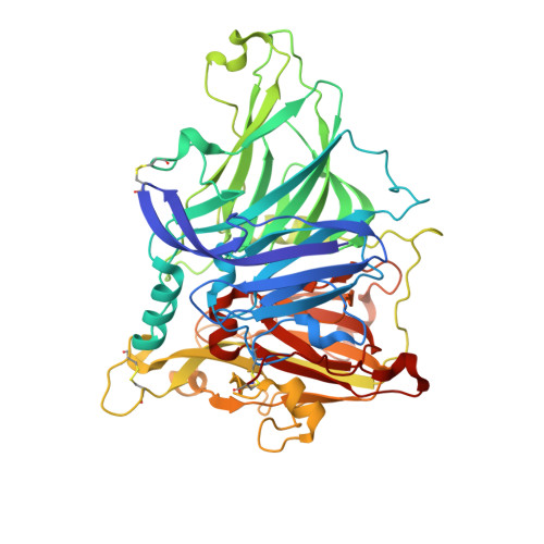
    Plant laccases catalyse the oxidation of monolignols in lignification, a process reinforcing the cell wall of many different cell types that provide mechanical support, nutrient transportation and defence against pathogens in plants 1 . The isozymes display a broad range of substrate preferences. Here, the substrate preference of a laccase (ZmLac3) from Zea mays (maize) was characterized. The crystal structure of ZmLac3 revealed a compact and deep substrate-binding pocket, and the binding modes of sinapyl alcohol (SinA) and coniferyl alcohol (ConA) were solved. On the basis of structural data and kinetics analysis, we propose that the regionalization of polar and hydrophobic surfaces in the binding pocket of ZmLac3 is vital for defining the orientation of SinA/ConA binding. The extra methoxyl group in SinA makes substantial contributions to interactions between SinA and ZmLac3, which are absent in the ZmLac3-ConA complex. In summary, the polar and hydrophobic interactions between SinA/ConA and ZmLac3 determine the binding positions of the monolignols in ZmLac3. These results provide valuable insight about ZmLac3 catalysis and should aid industrial processes that use plant laccases.


  • Organizational Affiliation

    Key Laboratory of Environmental and Applied Microbiology, Chengdu Institute of Biology, Chinese Academy of Sciences, Chengdu, China.


Macromolecules
Find similar proteins by:  (by identity cutoff)  |  3D Structure
Entity ID: 1
MoleculeChains Sequence LengthOrganismDetailsImage
Laccase550Zea maysMutation(s): 0 
Gene Names: lac3
EC: 1.10.3.2
UniProt
Find proteins for Q2PAJ1 (Zea mays)
Explore Q2PAJ1 
Go to UniProtKB:  Q2PAJ1
Entity Groups  
Sequence Clusters30% Identity50% Identity70% Identity90% Identity95% Identity100% Identity
UniProt GroupQ2PAJ1
Sequence Annotations
Expand
  • Reference Sequence
Oligosaccharides

Help

Entity ID: 2
MoleculeChains Length2D Diagram Glycosylation3D Interactions
2-acetamido-2-deoxy-beta-D-glucopyranose-(1-4)-2-acetamido-2-deoxy-beta-D-glucopyranose
B, C
2N-Glycosylation
Glycosylation Resources
GlyTouCan:  G42666HT
GlyCosmos:  G42666HT
GlyGen:  G42666HT
Small Molecules
Ligands 5 Unique
IDChains Name / Formula / InChI Key2D Diagram3D Interactions
NAG
Query on NAG

Download Ideal Coordinates CCD File 
H [auth A],
I [auth A],
J [auth A],
K [auth A],
L [auth A]
2-acetamido-2-deoxy-beta-D-glucopyranose
C8 H15 N O6
OVRNDRQMDRJTHS-FMDGEEDCSA-N
CIY (Subject of Investigation/LOI)
Query on CIY

Download Ideal Coordinates CCD File 
M [auth A](2E)-3-(4-hydroxy-3-methoxyphenyl)prop-2-enal
C10 H10 O3
DKZBBWMURDFHNE-NSCUHMNNSA-N
GOL
Query on GOL

Download Ideal Coordinates CCD File 
N [auth A]GLYCEROL
C3 H8 O3
PEDCQBHIVMGVHV-UHFFFAOYSA-N
CU
Query on CU

Download Ideal Coordinates CCD File 
D [auth A],
E [auth A],
F [auth A],
G [auth A]
COPPER (II) ION
Cu
JPVYNHNXODAKFH-UHFFFAOYSA-N
OXY
Query on OXY

Download Ideal Coordinates CCD File 
O [auth A]OXYGEN MOLECULE
O2
MYMOFIZGZYHOMD-UHFFFAOYSA-N
Experimental Data & Validation

Experimental Data

  • Method: X-RAY DIFFRACTION
  • Resolution: 2.00 Å
  • R-Value Free: 0.205 
  • R-Value Work: 0.164 
  • R-Value Observed: 0.166 
  • Space Group: P 61 2 2
Unit Cell:
Length ( Å )Angle ( ˚ )
a = 111.848α = 90
b = 111.848β = 90
c = 208.185γ = 120
Software Package:
Software NamePurpose
HKL-2000data reduction
HKL-2000data scaling
PHASERphasing
PHENIXrefinement
PDB_EXTRACTdata extraction

Structure Validation

View Full Validation Report


Ligand Structure Quality Assessment 


Entry History 

Deposition Data

Revision History  (Full details and data files)

  • Version 1.0: 2020-01-01
    Type: Initial release
  • Version 1.1: 2020-03-11
    Changes: Data collection, Database references
  • Version 1.2: 2020-04-01
    Changes: Database references
  • Version 2.0: 2020-07-29
    Type: Remediation
    Reason: Carbohydrate remediation
    Changes: Atomic model, Data collection, Derived calculations, Structure summary
  • Version 2.1: 2023-11-22
    Changes: Data collection, Database references, Refinement description, Structure summary