6K5G

Structural and catalytic analysis of two diverse uridine phosphorylases in the oomycete Phytophthora capsici


Experimental Data Snapshot

  • Method: X-RAY DIFFRACTION
  • Resolution: 1.57 Å
  • R-Value Free: 0.217 
  • R-Value Work: 0.186 
  • R-Value Observed: 0.188 

wwPDB Validation   3D Report Full Report


Ligand Structure Quality Assessment 


This is version 1.2 of the entry. See complete history


Literature

Structural and catalytic analysis of two diverse uridine phosphorylases in Phytophthora capsici.

Yang, C.Li, J.Huang, Z.Zhang, X.Gao, X.Zhu, C.Morris, P.F.Zhang, X.

(2020) Sci Rep 10: 9051-9051

  • DOI: https://doi.org/10.1038/s41598-020-65935-9
  • Primary Citation of Related Structures:  
    6K5G, 6K5H, 6K5K, 6K8P

  • PubMed Abstract: 

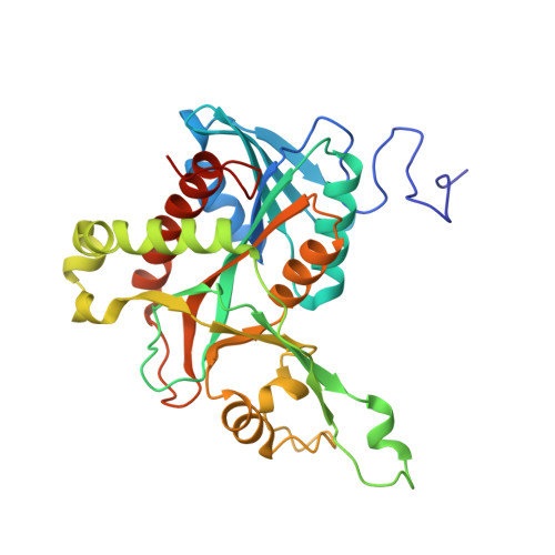
    Uridine phosphorylase (UP) is a key enzyme of pyrimidine salvage pathways that enables the recycling of endogenous or exogenous-supplied pyrimidines and plays an important intracellular metabolic role. Here, we biochemically and structurally characterized two evolutionarily divergent uridine phosphorylases, PcUP1 and PcUP2 from the oomycete pathogen Phytophthora capsici. Our analysis of other oomycete genomes revealed that both uridine phosphorylases are present in Phytophthora and Pythium genomes, but only UP2 is seen in Saprolegnia spp. which are basal members of the oomycetes. Moreover, uridine phosphorylases are not found in obligate oomycete pathogens such as Hyaloperonospora arabidopsidis and Albugo spp. PcUP1 and PcUP2 are upregulated 300 and 500 fold respectively, within 90 min after infection of pepper leaves. The crystal structures of PcUP1 in ligand-free and in complex with uracil/ribose-1-phosphate, 2'-deoxyuridine/phosphate and thymidine/phosphate were analyzed. Crystal structure of this uridine phosphorylase showed strict conservation of key residues in the binding pocket. Structure analysis of PcUP1 with bound ligands, and site-directed mutagenesis of key residues provide additional support for the "push-pull" model of catalysis. Our study highlights the importance of pyrimidine salvage during the earliest stages of infection.


  • Organizational Affiliation

    Shandong Provincial Key Laboratory for Biology of Vegetable Diseases and Insect Pests, College of Plant Protection, Shandong Agricultural University, Tai'an, 271000, China.


Macromolecules
Find similar proteins by:  (by identity cutoff)  |  3D Structure
Entity ID: 1
MoleculeChains Sequence LengthOrganismDetailsImage
Uridine phosphorylase
A, B, C, D
309Phytophthora capsici LT1534Mutation(s): 0 
Gene Names: up
EC: 2.4.2.3
UniProt
Find proteins for A0A410UCT3 (Phytophthora capsici)
Explore A0A410UCT3 
Go to UniProtKB:  A0A410UCT3
Entity Groups  
Sequence Clusters30% Identity50% Identity70% Identity90% Identity95% Identity100% Identity
UniProt GroupA0A410UCT3
Sequence Annotations
Expand
  • Reference Sequence
Experimental Data & Validation

Experimental Data

  • Method: X-RAY DIFFRACTION
  • Resolution: 1.57 Å
  • R-Value Free: 0.217 
  • R-Value Work: 0.186 
  • R-Value Observed: 0.188 
  • Space Group: P 1 21 1
Unit Cell:
Length ( Å )Angle ( ˚ )
a = 67.246α = 90
b = 153.058β = 115.75
c = 67.28γ = 90
Software Package:
Software NamePurpose
PHENIXrefinement
PDB_EXTRACTdata extraction
HKL-3000data reduction
HKL-3000data scaling
SOLVEphasing
BLU-MAXdata collection
PDB_EXTRACTdata extraction
RESOLVEphasing

Structure Validation

View Full Validation Report


Ligand Structure Quality Assessment 


Entry History & Funding Information

Deposition Data


Funding OrganizationLocationGrant Number
National Natural Science Foundation of ChinaChinaCARS-25-03B

Revision History  (Full details and data files)

  • Version 1.0: 2019-06-12
    Type: Initial release
  • Version 1.1: 2020-06-17
    Changes: Database references
  • Version 1.2: 2024-03-27
    Changes: Data collection, Database references