6JIX

The cyrstal structure of taurine:2-oxoglutarate aminotransferase from Bifidobacterium kashiwanohense, in complex with PLP and glutamate


Experimental Data Snapshot

  • Method: X-RAY DIFFRACTION
  • Resolution: 2.65 Å
  • R-Value Free: 0.227 
  • R-Value Work: 0.167 
  • R-Value Observed: 0.169 

wwPDB Validation   3D Report Full Report


Ligand Structure Quality Assessment 


This is version 2.1 of the entry. See complete history


Literature

Biochemical and structural investigation of taurine:2-oxoglutarate aminotransferase fromBifidobacterium kashiwanohense.

Li, M.Wei, Y.Yin, J.Lin, L.Zhou, Y.Hua, G.Cao, P.Ang, E.L.Zhao, H.Yuchi, Z.Zhang, Y.

(2019) Biochem J 476: 1605-1619

  • DOI: https://doi.org/10.1042/BCJ20190206
  • Primary Citation of Related Structures:  
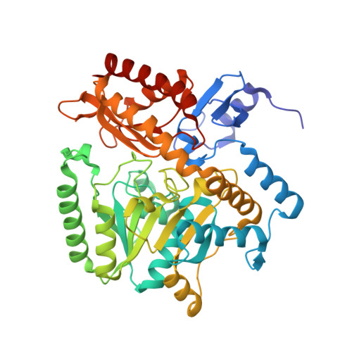
    6JIX

  • PubMed Abstract: 

    Taurine aminotransferases catalyze the first step in taurine catabolism in many taurine-degrading bacteria and play an important role in bacterial taurine metabolism in the mammalian gut. Here, we report the biochemical and structural characterization of a new taurine:2-oxoglutarate aminotransferase from the human gut bacterium Bifidobacterium kashiwanohense ( Bk Toa). Biochemical assays revealed high specificity of Bk Toa for 2-oxoglutarate as the amine acceptor. The crystal structure of Bk Toa in complex with pyridoxal 5'-phosphate (PLP) and glutamate was determined at 2.7 Å resolution. The enzyme forms a homodimer, with each monomer exhibiting a typical type I PLP-enzyme fold and conserved PLP-coordinating residues interacting with the PLP molecule. Two glutamate molecules are bound in sites near the predicted active site and they may occupy a path for substrate entry and product release. Molecular docking reveals a role for active site residues Trp21 and Arg156, conserved in Toa enzymes studied to date, in interacting with the sulfonate group of taurine. Bioinformatics analysis shows that the close homologs of Bk Toa are also present in other anaerobic gut bacteria.


  • Organizational Affiliation

    Tianjin Key Laboratory for Modern Drug Delivery & High-Efficiency, Collaborative Innovation Center of Chemical Science and Engineering, School of Pharmaceutical Science and Technology, Tianjin University, Tianjin 300072, China.


Macromolecules
Find similar proteins by:  (by identity cutoff)  |  3D Structure
Entity ID: 1
MoleculeChains Sequence LengthOrganismDetailsImage
taurine:2-oxoglutarate aminotransferaseA [auth B],
B [auth A],
C,
D
449Bifidobacterium catenulatum PV20-2Mutation(s): 0 
Gene Names: AH68_00255
UniProt
Find proteins for A0A0A7I435 (Bifidobacterium catenulatum PV20-2)
Explore A0A0A7I435 
Go to UniProtKB:  A0A0A7I435
Entity Groups  
Sequence Clusters30% Identity50% Identity70% Identity90% Identity95% Identity100% Identity
UniProt GroupA0A0A7I435
Sequence Annotations
Expand
  • Reference Sequence
Small Molecules
Ligands 2 Unique
IDChains Name / Formula / InChI Key2D Diagram3D Interactions
PLP
Query on PLP

Download Ideal Coordinates CCD File 
G [auth B],
I [auth A],
M [auth C],
P [auth D]
PYRIDOXAL-5'-PHOSPHATE
C8 H10 N O6 P
NGVDGCNFYWLIFO-UHFFFAOYSA-N
GLU
Query on GLU

Download Ideal Coordinates CCD File 
E [auth B]
F [auth B]
H [auth A]
J [auth C]
K [auth C]
E [auth B],
F [auth B],
H [auth A],
J [auth C],
K [auth C],
L [auth C],
N [auth D],
O [auth D]
GLUTAMIC ACID
C5 H9 N O4
WHUUTDBJXJRKMK-VKHMYHEASA-N
Experimental Data & Validation

Experimental Data

  • Method: X-RAY DIFFRACTION
  • Resolution: 2.65 Å
  • R-Value Free: 0.227 
  • R-Value Work: 0.167 
  • R-Value Observed: 0.169 
  • Space Group: P 1 21 1
Unit Cell:
Length ( Å )Angle ( ˚ )
a = 68.39α = 90
b = 164.544β = 105.81
c = 109.24γ = 90
Software Package:
Software NamePurpose
PHENIXrefinement
HKL-3000data scaling
PDB_EXTRACTdata extraction
HKL-3000data reduction
PHENIXphasing

Structure Validation

View Full Validation Report


Ligand Structure Quality Assessment 


Entry History & Funding Information

Deposition Data


Funding OrganizationLocationGrant Number
National Science Foundation (China)China31870049

Revision History  (Full details and data files)

  • Version 1.0: 2020-01-01
    Type: Initial release
  • Version 2.0: 2023-02-15
    Changes: Advisory, Atomic model, Data collection, Database references, Derived calculations, Non-polymer description, Structure summary
  • Version 2.1: 2023-11-29
    Changes: Data collection, Refinement description