


Crystal structure of SETD3 bound to Actin peptide and SFG
Li, H., Zheng, Y.To be published.
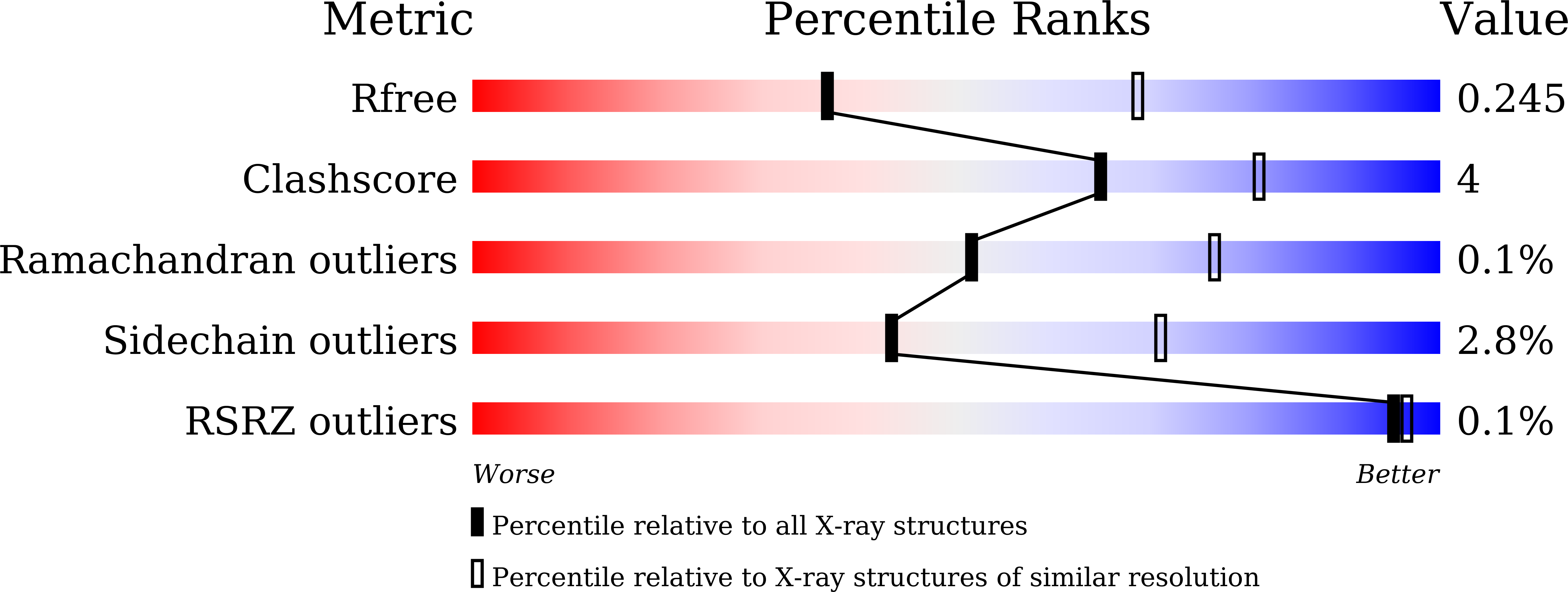
Experimental Data Snapshot
Entity ID: 1 | |||||
|---|---|---|---|---|---|
| Molecule | Chains | Sequence Length | Organism | Details | Image |
| Histone-lysine N-methyltransferase setd3 | 499 | Homo sapiens | Mutation(s): 0 Gene Names: SETD3, C14orf154 EC: 2.1.1.43 | 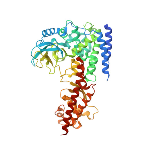 | |
UniProt & NIH Common Fund Data Resources | |||||
Find proteins for Q86TU7 (Homo sapiens) Explore Q86TU7 Go to UniProtKB: Q86TU7 | |||||
PHAROS: Q86TU7 GTEx: ENSG00000183576 | |||||
Entity Groups | |||||
| Sequence Clusters | 30% Identity50% Identity70% Identity90% Identity95% Identity100% Identity | ||||
| UniProt Group | Q86TU7 | ||||
Sequence AnnotationsExpand | |||||
| |||||
Find similar proteins by: Sequence | 3D Structure
Entity ID: 2 | |||||
|---|---|---|---|---|---|
| Molecule | Chains | Sequence Length | Organism | Details | Image |
| Actin, gamma-enteric smooth muscle | 22 | Homo sapiens | Mutation(s): 0 |  | |
UniProt & NIH Common Fund Data Resources | |||||
Find proteins for P63267 (Homo sapiens) Explore P63267 Go to UniProtKB: P63267 | |||||
PHAROS: P63267 GTEx: ENSG00000163017 | |||||
Entity Groups | |||||
| Sequence Clusters | 30% Identity50% Identity70% Identity90% Identity95% Identity100% Identity | ||||
| UniProt Group | P63267 | ||||
Sequence AnnotationsExpand | |||||
| |||||
| Ligands 3 Unique | |||||
|---|---|---|---|---|---|
| ID | Chains | Name / Formula / InChI Key | 2D Diagram | 3D Interactions | |
| SFG (Subject of Investigation/LOI) Query on SFG | E [auth A], I [auth C] | SINEFUNGIN C15 H23 N7 O5 LMXOHSDXUQEUSF-YECHIGJVSA-N |  | ||
| EPE Query on EPE | L [auth C] | 4-(2-HYDROXYETHYL)-1-PIPERAZINE ETHANESULFONIC ACID C8 H18 N2 O4 S JKMHFZQWWAIEOD-UHFFFAOYSA-N |  | ||
| SO4 Query on SO4 | F [auth A], G [auth A], H [auth A], J [auth C], K [auth C] | SULFATE ION O4 S QAOWNCQODCNURD-UHFFFAOYSA-L |  | ||
| Length ( Å ) | Angle ( ˚ ) |
|---|---|
| a = 60.434 | α = 90 |
| b = 131.282 | β = 90 |
| c = 175.153 | γ = 90 |
| Software Name | Purpose |
|---|---|
| DENZO | data reduction |
| SCALEPACK | data scaling |
| MOLREP | phasing |
| PHENIX | refinement |
| PDB_EXTRACT | data extraction |
| Funding Organization | Location | Grant Number |
|---|---|---|
| National Science Foundation (China) | China | 91753203 |
RCSB PDB (citation) is hosted by
RCSB PDB is a member of the

