6J8F

Crystal structure of SVBP-VASH1 with peptide mimic the C-terminal of alpha-tubulin


Experimental Data Snapshot

  • Method: X-RAY DIFFRACTION
  • Resolution: 2.28 Å
  • R-Value Free: 0.237 
  • R-Value Work: 0.200 
  • R-Value Observed: 0.202 

wwPDB Validation   3D Report Full Report


This is version 1.3 of the entry. See complete history


Literature

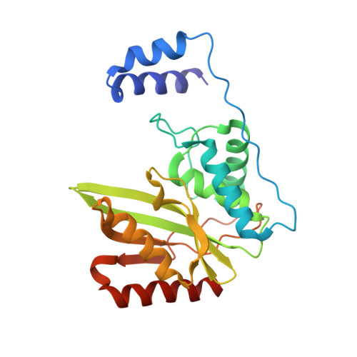
Molecular basis of vasohibins-mediated detyrosination and its impact on spindle function and mitosis.

Liao, S.Rajendraprasad, G.Wang, N.Eibes, S.Gao, J.Yu, H.Wu, G.Tu, X.Huang, H.Barisic, M.Xu, C.

(2019) Cell Res 29: 533-547

  • DOI: https://doi.org/10.1038/s41422-019-0187-y
  • Primary Citation of Related Structures:  
    6J7B, 6J8F, 6J8N, 6J91, 6J9H

  • PubMed Abstract: 

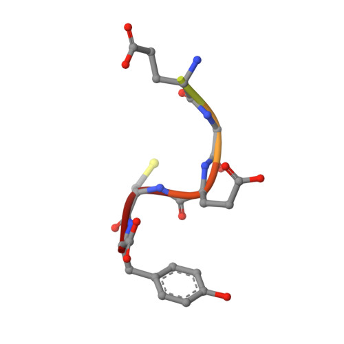
    α-Tubulin detyrosination, largely catalyzed by vasohibins, is involved in many microtubule (MT)-related cellular events. In this study, we identified a core heterodimeric complex of human small vasohibin-binding protein (SVBP) and vasohibin 1 (VASH1) (hereafter denoted as SVBP-VASH1) that catalyzes the detyrosination of a peptide derived from C-terminus of α-tubulin. We further solved the crystal structures of the SVBP-VASH1 heterodimer alone and in complex with either an inhibitor or a mutant substrate peptide. Our structural research, complemented by biochemical and mutagenesis experiments, resulted in identification of the key residues for VASH1 binding to SVBP and α-tubulin substrate. Our in vivo experiments reveal that MT detyrosination in general, as well as the interactions between SVBP, VASH1, and α-tubulin, are critical for spindle function and accurate chromosome segregation during mitosis. Furthermore, we found that the phenotypes caused by the depletion of vasohibins were largely rescued upon co-depletion of kinesin13/MCAK, suggesting the coordination between the MT depolymerase and MT detyrosination during mitosis. Thus our work not only provides structural insights into the molecular mechanism of α-tubulin detyrosination catalyzed by SVBP-bound vasohibins, but also uncovers the key role of vasohibins-mediated MT detyrosination in spindle morphology and chromosome segregation during mitosis.


  • Organizational Affiliation

    Hefei National Laboratory for Physical Sciences at Microscale and School of Life Sciences, University of Science and Technology of China, Hefei, China.


Macromolecules
Find similar proteins by:  (by identity cutoff)  |  3D Structure
Entity ID: 1
MoleculeChains Sequence LengthOrganismDetailsImage
Small vasohibin-binding protein46Homo sapiensMutation(s): 0 
Gene Names: SVBPCCDC23
UniProt & NIH Common Fund Data Resources
Find proteins for Q8N300 (Homo sapiens)
Explore Q8N300 
Go to UniProtKB:  Q8N300
PHAROS:  Q8N300
GTEx:  ENSG00000177868 
Entity Groups  
Sequence Clusters30% Identity50% Identity70% Identity90% Identity95% Identity100% Identity
UniProt GroupQ8N300
Sequence Annotations
Expand
  • Reference Sequence
Find similar proteins by:  (by identity cutoff)  |  3D Structure
Entity ID: 2
MoleculeChains Sequence LengthOrganismDetailsImage
Tubulinyl-Tyr carboxypeptidase 1238Homo sapiensMutation(s): 0 
Gene Names: VASH1KIAA1036VASH
EC: 3.4.17.17
UniProt & NIH Common Fund Data Resources
Find proteins for Q7L8A9 (Homo sapiens)
Explore Q7L8A9 
Go to UniProtKB:  Q7L8A9
PHAROS:  Q7L8A9
GTEx:  ENSG00000071246 
Entity Groups  
Sequence Clusters30% Identity50% Identity70% Identity90% Identity95% Identity100% Identity
UniProt GroupQ7L8A9
Sequence Annotations
Expand
  • Reference Sequence

Find similar proteins by:  Sequence   |   3D Structure  

Entity ID: 3
MoleculeChains Sequence LengthOrganismDetailsImage
8-mer peptide8Homo sapiensMutation(s): 0 
UniProt & NIH Common Fund Data Resources
Find proteins for Q71U36 (Homo sapiens)
Explore Q71U36 
Go to UniProtKB:  Q71U36
PHAROS:  Q71U36
GTEx:  ENSG00000167552 
Entity Groups  
UniProt GroupQ71U36
Sequence Annotations
Expand
  • Reference Sequence
Experimental Data & Validation

Experimental Data

  • Method: X-RAY DIFFRACTION
  • Resolution: 2.28 Å
  • R-Value Free: 0.237 
  • R-Value Work: 0.200 
  • R-Value Observed: 0.202 
  • Space Group: P 21 21 2
Unit Cell:
Length ( Å )Angle ( ˚ )
a = 66.95α = 90
b = 108.377β = 90
c = 46.896γ = 90
Software Package:
Software NamePurpose
PHENIXrefinement
XDSdata reduction
XDSdata scaling
PHENIXphasing

Structure Validation

View Full Validation Report


Entry History & Funding Information

Deposition Data


Funding OrganizationLocationGrant Number
National Natural Science Foundation of ChinaChina31500601
National Natural Science Foundation of ChinaChina31570737
National Natural Science Foundation of ChinaChina31770806

Revision History  (Full details and data files)

  • Version 1.0: 2019-06-19
    Type: Initial release
  • Version 1.1: 2019-07-17
    Changes: Data collection, Database references
  • Version 1.2: 2024-03-27
    Changes: Data collection, Database references
  • Version 1.3: 2024-04-03
    Changes: Refinement description