6IYQ

Crystal structure of a DNA duplex cross-linked by 6-thioguanine-6-thioguanine disulfides


Experimental Data Snapshot

  • Method: X-RAY DIFFRACTION
  • Resolution: 2.01 Å
  • R-Value Free: 0.282 
  • R-Value Work: 0.247 
  • R-Value Observed: 0.251 

wwPDB Validation   3D Report Full Report


This is version 1.1 of the entry. See complete history


Literature

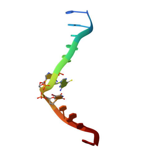
Crystal structure of a DNA duplex cross-linked by 6-thioguanine-6-thioguanine disulfides: reversible formation and cleavage catalyzed by Cu(II) ion and glutathione

Ono, A.Atsugi, T.Goto, M.Saneyoshi, H.Tomori, T.Seio, K.Dairaku, T.Kondo, J.

(2019) RSC Adv 


Macromolecules

Find similar nucleic acids by:  Sequence   |   3D Structure  

Entity ID: 1
MoleculeChains LengthOrganismImage
DNA (5'-D(*CP*GP*CP*GP*AP*(S6G)P*(S6G)P*(BRU)P*CP*GP*CP*G)-3')
A, B
12synthetic construct
Sequence Annotations
Expand
  • Reference Sequence
Experimental Data & Validation

Experimental Data

  • Method: X-RAY DIFFRACTION
  • Resolution: 2.01 Å
  • R-Value Free: 0.282 
  • R-Value Work: 0.247 
  • R-Value Observed: 0.251 
  • Space Group: I 2 2 2
Unit Cell:
Length ( Å )Angle ( ˚ )
a = 45.254α = 90
b = 46.522β = 90
c = 76.084γ = 90
Software Package:
Software NamePurpose
XSCALEdata scaling
SOLVEphasing
PHENIXrefinement
PDB_EXTRACTdata extraction
XDSdata reduction

Structure Validation

View Full Validation Report


Entry History 

Deposition Data

Revision History  (Full details and data files)

  • Version 1.0: 2019-10-30
    Type: Initial release
  • Version 1.1: 2024-03-27
    Changes: Data collection, Database references