6IUH

Crystal structure of GIT1 PBD domain in complex with Liprin-alpha2


Experimental Data Snapshot

  • Method: X-RAY DIFFRACTION
  • Resolution: 1.80 Å
  • R-Value Free: 0.267 
  • R-Value Work: 0.227 
  • R-Value Observed: 0.229 

wwPDB Validation   3D Report Full Report


This is version 1.2 of the entry. See complete history


Literature

Structural basis of the target-binding mode of the G protein-coupled receptor kinase-interacting protein in the regulation of focal adhesion dynamics.

Liang, M.Xie, X.Pan, J.Jin, G.Yu, C.Wei, Z.

(2019) J Biol Chem 294: 5827-5839

  • DOI: https://doi.org/10.1074/jbc.RA118.006915
  • Primary Citation of Related Structures:  
    6IUH, 6IUI

  • PubMed Abstract: 

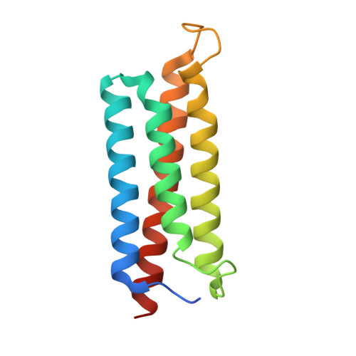
    Focal adhesions (FAs) are specialized sites where intracellular cytoskeleton elements connect to the extracellular matrix and thereby control cell motility. FA assembly depends on various scaffold proteins, including the G protein-coupled receptor kinase-interacting protein 1 (GIT1), paxillin, and liprin-α. Although liprin-α and paxillin are known to competitively interact with GIT1, the molecular basis governing these interactions remains elusive. To uncover the underlying mechanisms of how GIT1 is involved in FA assembly by alternatively binding to liprin-α and paxillin, here we solved the crystal structures of GIT1 in complex with liprin-α and paxillin at 1.8 and 2.6 Å resolutions, respectively. These structures revealed that the paxillin-binding domain (PBD) of GIT1 employs distinct binding modes to recognize a single α-helix of liprin-α and the LD4 motif of paxillin. Structure-based design of protein variants produced two binding-deficient GIT1 variants; specifically, these variants lost the ability to interact with liprin-α only or with both liprin-α and paxillin. Expressing the GIT1 variants in COS7 cells, we discovered that the two PBD-meditated interactions play different roles in either recruiting GIT1 to FA or facilitating FA assembly. Additionally, we demonstrate that, unlike for the known binding mode of the FAT domain to LD motifs, the PBD of GIT1 uses different surface patches to achieve high selectivity in LD motif recognition. In summary, our results have uncovered the mechanisms by which GIT1's PBD recognizes cognate paxillin and liprin-α structures, information we anticipate will be useful for future investigations of GIT1-protein interactions in cells.


  • Organizational Affiliation

    From the Department of Biology, Southern University of Science and Technology, Shenzhen 518055, China.


Macromolecules
Find similar proteins by:  (by identity cutoff)  |  3D Structure
Entity ID: 1
MoleculeChains Sequence LengthOrganismDetailsImage
ARF GTPase-activating protein GIT1
A, B
132Rattus norvegicusMutation(s): 0 
Gene Names: Git1
UniProt
Find proteins for Q9Z272 (Rattus norvegicus)
Explore Q9Z272 
Go to UniProtKB:  Q9Z272
Entity Groups  
Sequence Clusters30% Identity50% Identity70% Identity90% Identity95% Identity100% Identity
UniProt GroupQ9Z272
Sequence Annotations
Expand
  • Reference Sequence
Find similar proteins by:  (by identity cutoff)  |  3D Structure
Entity ID: 2
MoleculeChains Sequence LengthOrganismDetailsImage
Liprin-alpha-2
C, D
36Homo sapiensMutation(s): 0 
Gene Names: PPFIA2
UniProt & NIH Common Fund Data Resources
Find proteins for O75334 (Homo sapiens)
Explore O75334 
Go to UniProtKB:  O75334
PHAROS:  O75334
GTEx:  ENSG00000139220 
Entity Groups  
Sequence Clusters30% Identity50% Identity70% Identity90% Identity95% Identity100% Identity
UniProt GroupO75334
Sequence Annotations
Expand
  • Reference Sequence
Experimental Data & Validation

Experimental Data

  • Method: X-RAY DIFFRACTION
  • Resolution: 1.80 Å
  • R-Value Free: 0.267 
  • R-Value Work: 0.227 
  • R-Value Observed: 0.229 
  • Space Group: P 21 21 21
Unit Cell:
Length ( Å )Angle ( ˚ )
a = 88.2α = 90
b = 38.617β = 90
c = 99.134γ = 90
Software Package:
Software NamePurpose
PHENIXrefinement
HKL-3000data scaling
PDB_EXTRACTdata extraction
HKL-3000data reduction
PHASERphasing

Structure Validation

View Full Validation Report


Entry History & Funding Information

Deposition Data


Funding OrganizationLocationGrant Number
National Natural Science Foundation of ChinaChina31770791
National Natural Science Foundation of ChinaChina31570741

Revision History  (Full details and data files)

  • Version 1.0: 2019-02-20
    Type: Initial release
  • Version 1.1: 2019-04-24
    Changes: Data collection, Database references
  • Version 1.2: 2023-11-22
    Changes: Data collection, Database references, Refinement description