Crystal structure and biochemical study on argininosuccinate lyase from Mycobacterium tuberculosis.
Chen, X.B., Chen, J., Zhang, W., Wang, H., Liu, X., Zhou, W., Yang, H., Rao, Z.(2019) Biochem Biophys Res Commun 510: 116-121
- PubMed: 30665717
- DOI: https://doi.org/10.1016/j.bbrc.2019.01.061
- Primary Citation of Related Structures:
6IG5, 6IGA - PubMed Abstract:
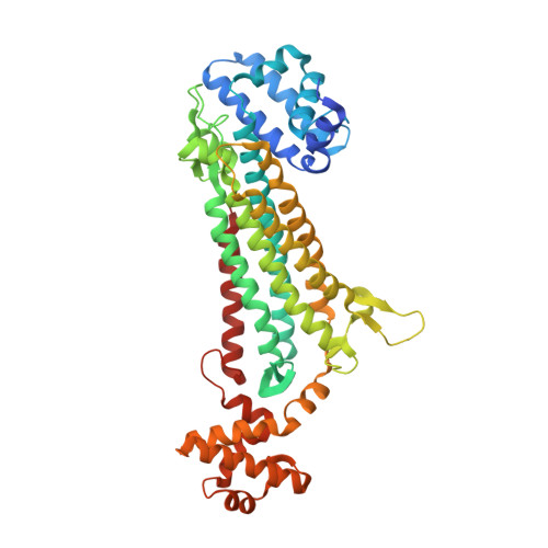
Argininosuccinate lyase (ASL) participates in arginine synthesis through catalysing a reversible reaction in which argininosuccinate (AS) converts into arginine and fumarate. ASL from Mycobacterium tuberculosis is essential for its growth. In this work, the crystal structure of the apo form of MtbASL was determined and reveals a tetrameric structure that is essential for its activity since the active sites are formed by residues from three different monomers. Subsequently, we determined the crystal structure of MtbASL-sulfate complex, and the ligand mimics the negatively charged intermediate. The complex structure and mutagenesis studies indicate that residues S282 and H161 might act as a catalytic dyad. A major conformational change in the MtbASL-SO 4 complex structure could be observed upon sulfate binding, and this movement facilitates the interaction between substrate and the residues involved in catalysis. A different conformational change in the C-terminal domain could be observed in the MtbASL-SO 4 complex compared with that in other homologues. This difference may be responsible for the lower activity of MtbASL, which is related to the slow growth rate of M. tuberculosis. The C-terminal domain is a potential allosteric site upon inhibitor binding. The various conformational changes and the diversity of the sequence of the potential allosteric site across the homologues might provide clues for designing selective inhibitors against M. tuberculosis.
Organizational Affiliation:
State Key Laboratory of Medicinal Chemical Biology and College of Pharmacy, Nankai University, Tianjin, China; College of Life Sciences, Nankai University, Tianjin, China.