6IF4

Crystal structure of Tbtudor


Experimental Data Snapshot

  • Method: X-RAY DIFFRACTION
  • Resolution: 1.93 Å
  • R-Value Free: 0.249 
  • R-Value Work: 0.206 
  • R-Value Observed: 0.208 

wwPDB Validation   3D Report Full Report


This is version 2.2 of the entry. See complete history


Literature

Crystal structure of TbEsa1 presumed Tudor domain from Trypanosoma brucei.

Gao, J.Ye, K.Diwu, Y.Xu, C.Zhang, X.Liao, S.Tu, X.

(2020) J Struct Biol 209: 107406-107406

  • DOI: https://doi.org/10.1016/j.jsb.2019.107406
  • Primary Citation of Related Structures:  
    6IF4

  • PubMed Abstract: 

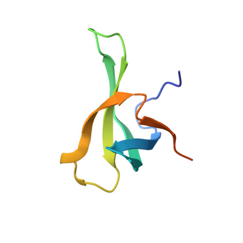
    The essential SAS2-related acetyltransferase 1 (Esa1), as a acetyltransferase of MYST family, is indispensable for the cell cycle and transcriptional regulation. The Tudor domain consists of 60 amino acids and belongs to the Royal family, which serves as a module interacting with methylated histone and/or DNA. Although Tudor domain has been widely studied in higher eukaryotes, its structure and function remain unclear in Trypanosoma brucei (T. brucei), a protozoan unicellular parasite causing sleeping sickness in human and nagana in cattle in sub-Saharan Africa. Here, we determined a high-resolution structure of TbEsa1 presumed Tudor domain from T. brucei by X-ray crystallography. TbEsa1 Tudor domain adopts a conserved Tudor-like fold, which is comprised of a five-stranded β-barrel surrounded by two short α-helices. Furthermore, we revealed a non-specific DNA binding pattern of TbEsa1 Tudor domain. However, TbEsa1 Tudor domain showed no methyl-histone binding ability, due to the absence of key aromatic residues forming a conserved aromatic cage.


  • Organizational Affiliation

    Hefei National Laboratory for Physical Sciences at Microscale, and School of Life Sciences, University of Science and Technology of China, Hefei, Anhui, PR China.


Macromolecules
Find similar proteins by:  (by identity cutoff)  |  3D Structure
Entity ID: 1
MoleculeChains Sequence LengthOrganismDetailsImage
Histone acetyltransferaseA [auth B],
B [auth A]
71Trypanosoma brucei brucei TREU927Mutation(s): 0 
Gene Names: Tb927.7.4560
EC: 2.3.1.48
UniProt
Find proteins for D6XIY7 (Trypanosoma brucei brucei (strain 927/4 GUTat10.1))
Explore D6XIY7 
Go to UniProtKB:  D6XIY7
Entity Groups  
Sequence Clusters30% Identity50% Identity70% Identity90% Identity95% Identity100% Identity
UniProt GroupD6XIY7
Sequence Annotations
Expand
  • Reference Sequence
Experimental Data & Validation

Experimental Data

  • Method: X-RAY DIFFRACTION
  • Resolution: 1.93 Å
  • R-Value Free: 0.249 
  • R-Value Work: 0.206 
  • R-Value Observed: 0.208 
  • Space Group: P 1
Unit Cell:
Length ( Å )Angle ( ˚ )
a = 29.389α = 113.58
b = 34.274β = 100.53
c = 37.47γ = 102.9
Software Package:
Software NamePurpose
PHENIXrefinement
XDSdata reduction
XDSdata scaling
PHENIXphasing

Structure Validation

View Full Validation Report


Entry History & Funding Information

Deposition Data


Funding OrganizationLocationGrant Number
National Natural Science Foundation of ChinaChina31500601
National Natural Science Foundation of ChinaChinaU1332137
National Natural Science Foundation of ChinaChina31570737

Revision History  (Full details and data files)

  • Version 1.0: 2019-09-18
    Type: Initial release
  • Version 2.0: 2019-11-06
    Type: Coordinate replacement
    Reason: Atomic clashes
    Changes: Advisory, Atomic model, Data collection, Derived calculations, Other, Refinement description, Structure summary
  • Version 2.1: 2020-09-30
    Changes: Database references
  • Version 2.2: 2023-11-22
    Changes: Data collection, Database references, Refinement description