6I8N

Crystal structure of LmrR with V15 replaced by unnatural amino acid 4-amino-L-phenylalanine


Experimental Data Snapshot

  • Method: X-RAY DIFFRACTION
  • Resolution: 1.79 Å
  • R-Value Free: 0.195 
  • R-Value Work: 0.172 
  • R-Value Observed: 0.173 

wwPDB Validation   3D Report Full Report


Ligand Structure Quality Assessment 


This is version 1.2 of the entry. See complete history


Literature

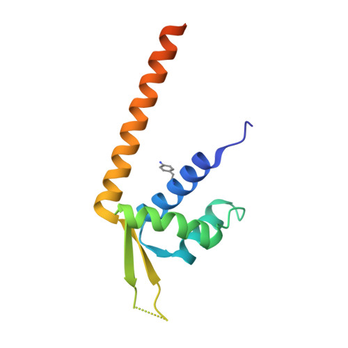
Directed Evolution of a Designer Enzyme Featuring an Unnatural Catalytic Amino Acid.

Mayer, C.Dulson, C.Reddem, E.Thunnissen, A.W.H.Roelfes, G.

(2019) Angew Chem Int Ed Engl 58: 2083-2087

  • DOI: https://doi.org/10.1002/anie.201813499
  • Primary Citation of Related Structures:  
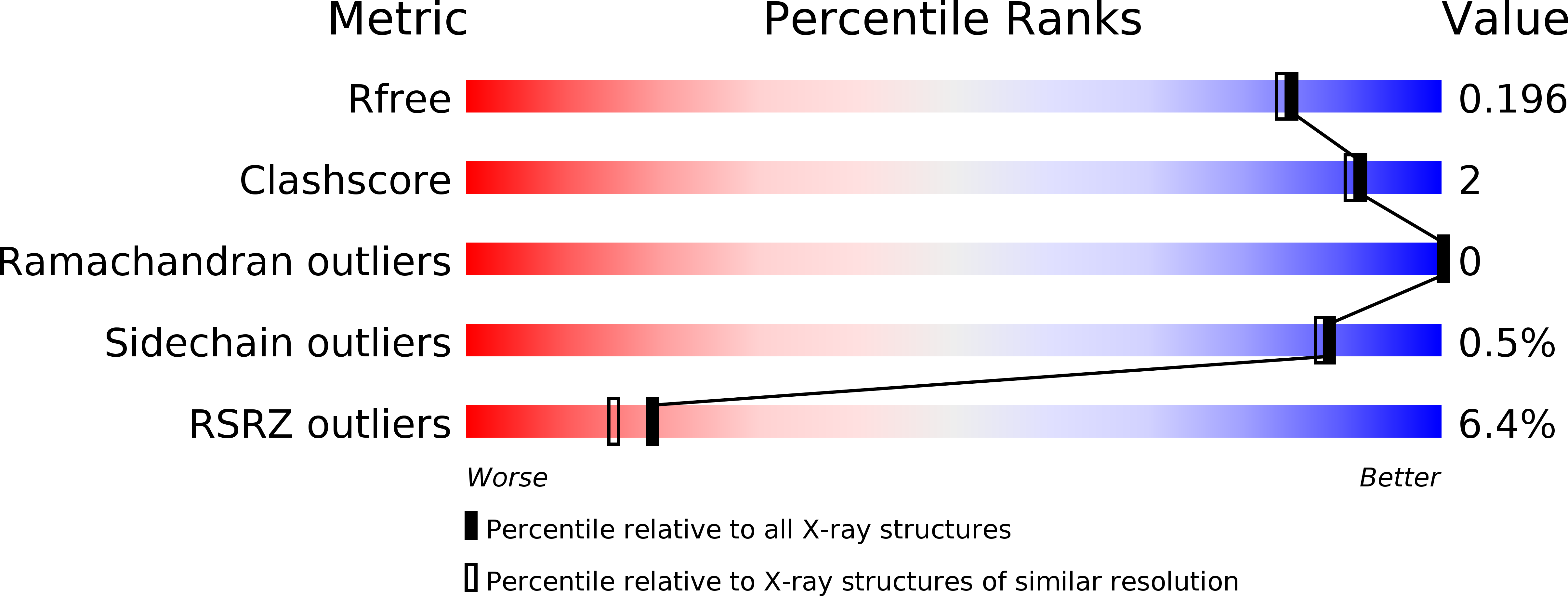
    6I8N

  • PubMed Abstract: 

    The impressive rate accelerations that enzymes display in nature often result from boosting the inherent catalytic activities of side chains by their precise positioning inside a protein binding pocket. Such fine-tuning is also possible for catalytic unnatural amino acids. Specifically, the directed evolution of a recently described designer enzyme, which utilizes an aniline side chain to promote a model hydrazone formation reaction, is reported. Consecutive rounds of directed evolution identified several mutations in the promiscuous binding pocket, in which the unnatural amino acid is embedded in the starting catalyst. When combined, these mutations boost the turnover frequency (k cat ) of the designer enzyme by almost 100-fold. This results from strengthening the catalytic contribution of the unnatural amino acid, as the engineered designer enzymes outperform variants, in which the aniline side chain is replaced with a catalytically inactive tyrosine residue, by more than 200-fold.


  • Organizational Affiliation

    Stratingh Institute for Chemistry, University of Groningen, Nijenborgh 4, 9474, AG, Groningen, The Netherlands.


Macromolecules
Find similar proteins by:  (by identity cutoff)  |  3D Structure
Entity ID: 1
MoleculeChains Sequence LengthOrganismDetailsImage
Transcriptional regulator, PadR-like family
A, B
126Lactococcus cremoris subsp. cremoris MG1363Mutation(s): 1 
Gene Names: llmg_0323
UniProt
Find proteins for A2RI36 (Lactococcus lactis subsp. cremoris (strain MG1363))
Explore A2RI36 
Go to UniProtKB:  A2RI36
Entity Groups  
Sequence Clusters30% Identity50% Identity70% Identity90% Identity95% Identity100% Identity
UniProt GroupA2RI36
Sequence Annotations
Expand
  • Reference Sequence
Small Molecules
Ligands 1 Unique
IDChains Name / Formula / InChI Key2D Diagram3D Interactions
MPO
Query on MPO

Download Ideal Coordinates CCD File 
C [auth A]3[N-MORPHOLINO]PROPANE SULFONIC ACID
C7 H15 N O4 S
DVLFYONBTKHTER-UHFFFAOYSA-N
Modified Residues  1 Unique
IDChains TypeFormula2D DiagramParent
HOX
Query on HOX
A, B
L-PEPTIDE LINKINGC9 H12 N2 O2PHE
Experimental Data & Validation

Experimental Data

  • Method: X-RAY DIFFRACTION
  • Resolution: 1.79 Å
  • R-Value Free: 0.195 
  • R-Value Work: 0.172 
  • R-Value Observed: 0.173 
  • Space Group: P 21 21 21
Unit Cell:
Length ( Å )Angle ( ˚ )
a = 35.727α = 90
b = 55.149β = 90
c = 145.085γ = 90
Software Package:
Software NamePurpose
PHENIXrefinement
XDSdata reduction
Aimlessdata scaling
PDB_EXTRACTdata extraction
PHASERphasing

Structure Validation

View Full Validation Report


Ligand Structure Quality Assessment 


Entry History & Funding Information

Deposition Data


Funding OrganizationLocationGrant Number
Netherlands Organisation for Scientific ResearchNetherlands724.013.003

Revision History  (Full details and data files)

  • Version 1.0: 2019-01-02
    Type: Initial release
  • Version 1.1: 2019-02-13
    Changes: Data collection, Database references
  • Version 1.2: 2024-01-24
    Changes: Data collection, Database references, Refinement description