


CdaA/GlmM complex in Bacillus subtilis
Tosi, T., Hoshiga, F.To be published.
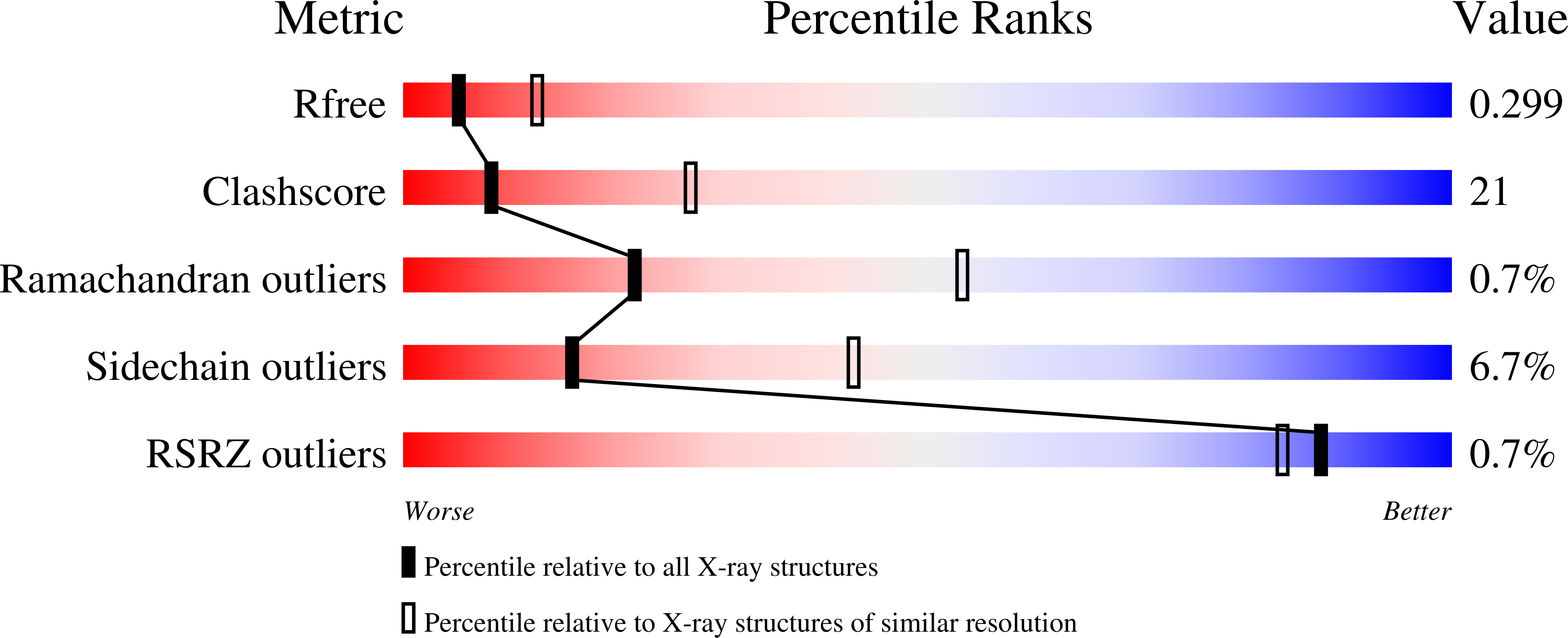
Experimental Data Snapshot
wwPDB Validation 3D Report Full Report
Entity ID: 1 | |||||
|---|---|---|---|---|---|
| Molecule | Chains | Sequence Length | Organism | Details | Image |
| Diadenylate cyclase | 183 | Bacillus subtilis | Mutation(s): 0 Gene Names: dacA, AX282_00750, B4417_1452, BS21228_13325, C7T97_16060, DJ572_00670, DLD52_20680, SC09_Contig25orf01072 EC: 2.7.7.85 |  | |
UniProt | |||||
Find proteins for Q45589 (Bacillus subtilis (strain 168)) Explore Q45589 Go to UniProtKB: Q45589 | |||||
Entity Groups | |||||
| Sequence Clusters | 30% Identity50% Identity70% Identity90% Identity95% Identity100% Identity | ||||
| UniProt Group | Q45589 | ||||
Sequence AnnotationsExpand | |||||
| |||||
| Length ( Å ) | Angle ( ˚ ) |
|---|---|
| a = 62.886 | α = 90 |
| b = 62.886 | β = 90 |
| c = 187.324 | γ = 90 |
| Software Name | Purpose |
|---|---|
| Aimless | data scaling |
| PHENIX | refinement |
| PDB_EXTRACT | data extraction |
| DIALS | data reduction |
| PHASER | phasing |
RCSB PDB (citation) is hosted by
RCSB PDB is a member of the

