6HTN

Structure of a fucose lectin from Kordia zhangzhouensis in complex with methyl-fucoside


Experimental Data Snapshot

  • Method: X-RAY DIFFRACTION
  • Resolution: 1.55 Å
  • R-Value Free: 0.180 
  • R-Value Work: 0.155 
  • R-Value Observed: 0.157 

wwPDB Validation   3D Report Full Report


Ligand Structure Quality Assessment 


This is version 1.2 of the entry. See complete history


Literature

Architecture and Evolution of Blade Assembly in beta-propeller Lectins.

Bonnardel, F.Kumar, A.Wimmerova, M.Lahmann, M.Perez, S.Varrot, A.Lisacek, F.Imberty, A.

(2019) Structure 27: 764-775.e3

  • DOI: https://doi.org/10.1016/j.str.2019.02.002
  • Primary Citation of Related Structures:  
    6HTN

  • PubMed Abstract: 

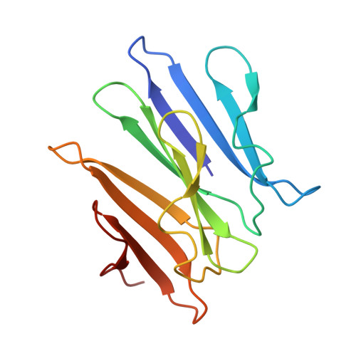
    Lectins with a β-propeller fold bind glycans on the cell surface through multivalent binding sites and appropriate directionality. These proteins are formed by repeats of short domains, raising questions about evolutionary duplication. However, these repeats are difficult to detect in translated genomes and seldom correctly annotated in sequence databases. To address these issues, we defined the blade signature of the five types of β-propellers using 3D-structural data. With these templates, we predicted 3,887 β-propeller lectins in 1,889 species and organized this information in a searchable online database. The data reveal a widespread distribution of β-propeller lectins across species. Prediction also emphasizes multiple architectures and led to the discovery of a β-propeller assembly scenario. This was confirmed by producing and characterizing a predicted protein coded in the genome of Kordia zhangzhouensis. The crystal structure uncovers an intermediate in the evolution of β-propeller assembly and demonstrates the power of our tools.


  • Organizational Affiliation

    University of Grenoble Alpes, CNRS, CERMAV, 38000 Grenoble, France; Swiss Institute of Bioinformatics, 1227 Geneva, Switzerland; Computer Science Department, UniGe, 1227 Geneva, Switzerland.


Macromolecules
Find similar proteins by:  (by identity cutoff)  |  3D Structure
Entity ID: 1
MoleculeChains Sequence LengthOrganismDetailsImage
Fucose-binding lectin
A, B, C, D, E
A, B, C, D, E, F
144Kordia periserrulaeMutation(s): 0 
Gene Names: C8N46_102337
UniProt
Find proteins for A0A2T6C3M6 (Kordia periserrulae)
Explore A0A2T6C3M6 
Go to UniProtKB:  A0A2T6C3M6
Entity Groups  
Sequence Clusters30% Identity50% Identity70% Identity90% Identity95% Identity100% Identity
UniProt GroupA0A2T6C3M6
Sequence Annotations
Expand
  • Reference Sequence
Small Molecules
Ligands 4 Unique
IDChains Name / Formula / InChI Key2D Diagram3D Interactions
PE8
Query on PE8

Download Ideal Coordinates CCD File 
Q [auth B]3,6,9,12,15,18,21-HEPTAOXATRICOSANE-1,23-DIOL
C16 H34 O9
GLZWNFNQMJAZGY-UHFFFAOYSA-N
MFU
Query on MFU

Download Ideal Coordinates CCD File 
BA [auth E]
CA [auth E]
DA [auth E]
G [auth A]
GA [auth F]
BA [auth E],
CA [auth E],
DA [auth E],
G [auth A],
GA [auth F],
H [auth A],
HA [auth F],
I [auth A],
IA [auth F],
M [auth B],
N [auth B],
O [auth B],
R [auth C],
S [auth C],
T [auth C],
X [auth D],
Y [auth D],
Z [auth D]
methyl alpha-L-fucopyranoside
C7 H14 O5
OHWCAVRRXKJCRB-CXNFULCWSA-N
MRD
Query on MRD

Download Ideal Coordinates CCD File 
AA [auth D]
EA [auth E]
J [auth A]
JA [auth F]
P [auth B]
AA [auth D],
EA [auth E],
J [auth A],
JA [auth F],
P [auth B],
U [auth C]
(4R)-2-METHYLPENTANE-2,4-DIOL
C6 H14 O2
SVTBMSDMJJWYQN-RXMQYKEDSA-N
EDO
Query on EDO

Download Ideal Coordinates CCD File 
FA [auth E],
K [auth A],
L [auth A],
V [auth C],
W [auth C]
1,2-ETHANEDIOL
C2 H6 O2
LYCAIKOWRPUZTN-UHFFFAOYSA-N
Binding Affinity Annotations 
IDSourceBinding Affinity
MFU Binding MOAD:  6HTN Kd: 860 (nM) from 1 assay(s)
Experimental Data & Validation

Experimental Data

  • Method: X-RAY DIFFRACTION
  • Resolution: 1.55 Å
  • R-Value Free: 0.180 
  • R-Value Work: 0.155 
  • R-Value Observed: 0.157 
  • Space Group: P 2 21 21
Unit Cell:
Length ( Å )Angle ( ˚ )
a = 87.102α = 90
b = 90.522β = 90
c = 143.777γ = 90
Software Package:
Software NamePurpose
REFMACrefinement
PDB_EXTRACTdata extraction
XDSdata reduction
Aimlessdata scaling
SHELXDphasing

Structure Validation

View Full Validation Report


Ligand Structure Quality Assessment 


Entry History & Funding Information

Deposition Data

  • Released Date: 2019-01-23 
  • Deposition Author(s): Varrot, A.

Funding OrganizationLocationGrant Number
French National Research AgencyFranceANR-15-IDEX-02

Revision History  (Full details and data files)

  • Version 1.0: 2019-01-23
    Type: Initial release
  • Version 1.1: 2019-08-07
    Changes: Data collection, Database references
  • Version 1.2: 2020-07-29
    Type: Remediation
    Reason: Carbohydrate remediation
    Changes: Data collection, Derived calculations, Structure summary