


Structural analysis of the PATZ1 BTB domain homodimer
Piepoli, S., Alt, A., Atilgan, C., Mancini, E.J., Erman, B.(2020) Acta Crystallogr D Biol Crystallogr
Experimental Data Snapshot
wwPDB Validation 3D Report Full Report

(2020) Acta Crystallogr D Biol Crystallogr
Entity ID: 1 | |||||
|---|---|---|---|---|---|
| Molecule | Chains | Sequence Length | Organism | Details | Image |
| POZ (BTB) and AT hook-containing zinc finger 1 | 155 | Danio rerio | Mutation(s): 0 Gene Names: patz1 | 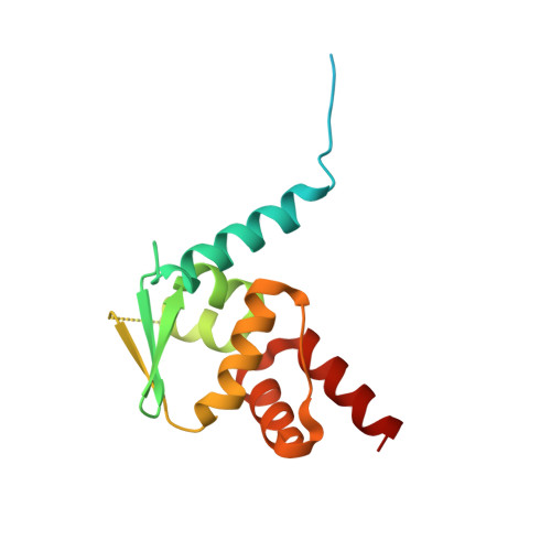 | |
UniProt | |||||
Find proteins for X1WGP9 (Danio rerio) Explore X1WGP9 Go to UniProtKB: X1WGP9 | |||||
Entity Groups | |||||
| Sequence Clusters | 30% Identity50% Identity70% Identity90% Identity95% Identity100% Identity | ||||
| UniProt Group | X1WGP9 | ||||
Sequence AnnotationsExpand | |||||
| |||||
| Length ( Å ) | Angle ( ˚ ) |
|---|---|
| a = 43.08 | α = 90 |
| b = 43.08 | β = 90 |
| c = 123.64 | γ = 120 |
| Software Name | Purpose |
|---|---|
| PHENIX | refinement |
| xia2 | data reduction |
| XDS | data scaling |
| PHASER | phasing |
| Funding Organization | Location | Grant Number |
|---|---|---|
| Royal Society | United Kingdom | NI140172 |
RCSB PDB (citation) is hosted by
RCSB PDB is a member of the

