6GMP

CRYSTAL STRUCTURE OF THE PPIASE DOMAIN OF TBPAR42


Experimental Data Snapshot

  • Method: X-RAY DIFFRACTION
  • Resolution: 1.35 Å
  • R-Value Free: 0.206 
  • R-Value Work: 0.166 
  • R-Value Observed: 0.168 

wwPDB Validation   3D Report Full Report


This is version 1.4 of the entry. See complete history


Literature

Structural Analysis of the 42 kDa Parvulin ofTrypanosoma brucei.

Rehic, E.Hoenig, D.Kamba, B.E.Goehring, A.Hofmann, E.Gasper, R.Matena, A.Bayer, P.

(2019) Biomolecules 9

  • DOI: https://doi.org/10.3390/biom9030093
  • Primary Citation of Related Structures:  
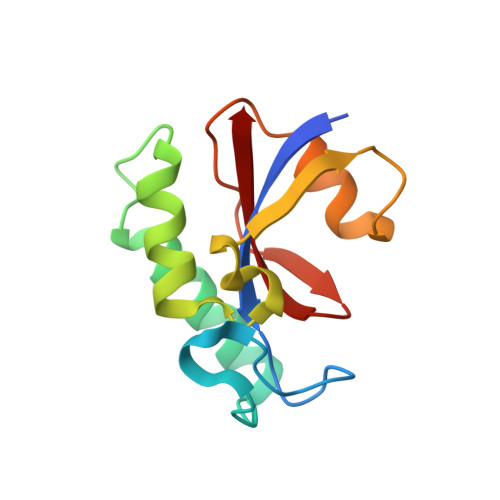
    2N84, 2N87, 6GMP

  • PubMed Abstract: 

    Trypanosoma brucei is a unicellular eukaryotic parasite, which causes the African sleeping sickness in humans. The recently discovered trypanosomal protein Parvulin 42 ( Tb Par42) plays a key role in parasite cell proliferation. Homologues of this two-domain protein are exclusively found in protozoa species. Tb Par42 exhibits an N-terminal forkhead associated (FHA)-domain and a peptidyl-prolyl- cis/trans -isomerase (PPIase) domain, both connected by a linker. Using NMR and X-ray analysis as well as activity assays, we report on the structures of the single domains of Tb Par42, discuss their intra-molecular interplay, and give some initial hints as to potential cellular functions of the protein.


  • Organizational Affiliation

    University Duisburg-Essen, Research Group Structural and Medicinal Biochemistry, Centre for Medical Biotechnology (ZMB), University of Duisburg-Essen, 45117 Essen, Germany. edisa.rehic@uni-due.de.


Macromolecules
Find similar proteins by:  (by identity cutoff)  |  3D Structure
Entity ID: 1
MoleculeChains Sequence LengthOrganismDetailsImage
PARVULIN 42122Trypanosoma brucei brucei TREU927Mutation(s): 0 
Gene Names: Tb927.7.2480
UniProt
Find proteins for Q57XM6 (Trypanosoma brucei brucei (strain 927/4 GUTat10.1))
Explore Q57XM6 
Go to UniProtKB:  Q57XM6
Entity Groups  
Sequence Clusters30% Identity50% Identity70% Identity90% Identity95% Identity100% Identity
UniProt GroupQ57XM6
Sequence Annotations
Expand
  • Reference Sequence
Experimental Data & Validation

Experimental Data

  • Method: X-RAY DIFFRACTION
  • Resolution: 1.35 Å
  • R-Value Free: 0.206 
  • R-Value Work: 0.166 
  • R-Value Observed: 0.168 
  • Space Group: C 2 2 21
Unit Cell:
Length ( Å )Angle ( ˚ )
a = 28.64α = 90
b = 69.96β = 90
c = 102.92γ = 90
Software Package:
Software NamePurpose
XDSdata scaling
XSCALEdata scaling
MOLREPphasing
PHENIXrefinement

Structure Validation

View Full Validation Report


Entry History & Funding Information

Deposition Data


Funding OrganizationLocationGrant Number
German Research FoundationGermanyResearch Training Group 1431

Revision History  (Full details and data files)

  • Version 1.0: 2019-03-20
    Type: Initial release
  • Version 1.1: 2019-03-27
    Changes: Data collection, Database references
  • Version 1.2: 2019-07-10
    Changes: Data collection, Database references
  • Version 1.3: 2019-07-17
    Changes: Data collection
  • Version 1.4: 2024-05-01
    Changes: Data collection, Database references, Refinement description