6GE9

Structure of Mycobacterium tuberculosis GlmU bound to Glc-1P and Ac-CoA


Experimental Data Snapshot

  • Method: X-RAY DIFFRACTION
  • Resolution: 2.26 Å
  • R-Value Free: 0.277 
  • R-Value Work: 0.243 
  • R-Value Observed: 0.245 

wwPDB Validation   3D Report Full Report


Ligand Structure Quality Assessment 


This is version 1.4 of the entry. See complete history


Literature

The Mechanism of Acetyl Transfer Catalyzed by Mycobacterium tuberculosis GlmU.

Craggs, P.D.Mouilleron, S.Rejzek, M.de Chiara, C.Young, R.J.Field, R.A.Argyrou, A.de Carvalho, L.P.S.

(2018) Biochemistry 57: 3387-3401

  • DOI: https://doi.org/10.1021/acs.biochem.8b00121
  • Primary Citation of Related Structures:  
    6GE9

  • PubMed Abstract: 

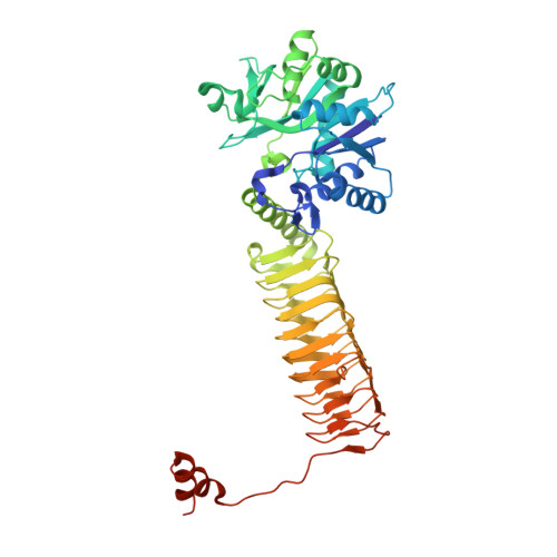
    The biosynthetic pathway of peptidoglycan is essential for Mycobacterium tuberculosis. We report here the acetyltransferase substrate specificity and catalytic mechanism of the bifunctional N-acetyltransferase/uridylyltransferase from M. tuberculosis (GlmU). This enzyme is responsible for the final two steps of the synthesis of UDP- N-acetylglucosamine, which is an essential precursor of peptidoglycan, from glucosamine 1-phosphate, acetyl-coenzyme A, and uridine 5'-triphosphate. GlmU utilizes ternary complex formation to transfer an acetyl from acetyl-coenzyme A to glucosamine 1-phosphate to form N-acetylglucosamine 1-phosphate. Steady-state kinetic studies and equilibrium binding experiments indicate that GlmU follows a steady-state ordered kinetic mechanism, with acetyl-coenzyme A binding first, which triggers a conformational change in GlmU, followed by glucosamine 1-phosphate binding. Coenzyme A is the last product to dissociate. Chemistry is partially rate-limiting as indicated by pH-rate studies and solvent kinetic isotope effects. A novel crystal structure of a mimic of the Michaelis complex, with glucose 1-phosphate and acetyl-coenzyme A, helps us to propose the residues involved in deprotonation of glucosamine 1-phosphate and the loop movement that likely generates the active site required for glucosamine 1-phosphate to bind. Together, these results pave the way for the rational discovery of improved inhibitors against M. tuberculosis GlmU, some of which might become candidates for antibiotic discovery programs.


  • Organizational Affiliation

    Platform Technology and Science , GlaxoSmithKline , Stevenage , U.K.


Macromolecules
Find similar proteins by:  (by identity cutoff)  |  3D Structure
Entity ID: 1
MoleculeChains Sequence LengthOrganismDetailsImage
Bifunctional protein GlmU495Mycobacterium tuberculosis H37RaMutation(s): 0 
Gene Names: glmUMRA_1026
EC: 2.7.7.23 (PDB Primary Data), 2.3.1.157 (PDB Primary Data)
UniProt
Find proteins for P9WMN3 (Mycobacterium tuberculosis (strain ATCC 25618 / H37Rv))
Explore P9WMN3 
Go to UniProtKB:  P9WMN3
Entity Groups  
Sequence Clusters30% Identity50% Identity70% Identity90% Identity95% Identity100% Identity
UniProt GroupP9WMN3
Sequence Annotations
Expand
  • Reference Sequence
Small Molecules
Ligands 5 Unique
IDChains Name / Formula / InChI Key2D Diagram3D Interactions
ACO
Query on ACO

Download Ideal Coordinates CCD File 
D [auth A]ACETYL COENZYME *A
C23 H38 N7 O17 P3 S
ZSLZBFCDCINBPY-ZSJPKINUSA-N
G1P
Query on G1P

Download Ideal Coordinates CCD File 
C [auth A]1-O-phosphono-alpha-D-glucopyranose
C6 H13 O9 P
HXXFSFRBOHSIMQ-VFUOTHLCSA-N
PEG
Query on PEG

Download Ideal Coordinates CCD File 
E [auth A],
F [auth A]
DI(HYDROXYETHYL)ETHER
C4 H10 O3
MTHSVFCYNBDYFN-UHFFFAOYSA-N
EDO
Query on EDO

Download Ideal Coordinates CCD File 
G [auth A],
H [auth A],
I [auth A]
1,2-ETHANEDIOL
C2 H6 O2
LYCAIKOWRPUZTN-UHFFFAOYSA-N
MG
Query on MG

Download Ideal Coordinates CCD File 
B [auth A]MAGNESIUM ION
Mg
JLVVSXFLKOJNIY-UHFFFAOYSA-N
Binding Affinity Annotations 
IDSourceBinding Affinity
ACO Binding MOAD:  6GE9 Kd: 2.50e+5 (nM) from 1 assay(s)
Experimental Data & Validation

Experimental Data

  • Method: X-RAY DIFFRACTION
  • Resolution: 2.26 Å
  • R-Value Free: 0.277 
  • R-Value Work: 0.243 
  • R-Value Observed: 0.245 
  • Space Group: H 3 2
Unit Cell:
Length ( Å )Angle ( ˚ )
a = 109.753α = 90
b = 109.753β = 90
c = 364.894γ = 120
Software Package:
Software NamePurpose
PHENIXrefinement
xia2data reduction
xia2data scaling
PHASERphasing

Structure Validation

View Full Validation Report


Ligand Structure Quality Assessment 


Entry History 

Deposition Data

Revision History  (Full details and data files)

  • Version 1.0: 2018-05-02
    Type: Initial release
  • Version 1.1: 2018-05-09
    Changes: Data collection, Database references
  • Version 1.2: 2018-06-27
    Changes: Data collection, Database references
  • Version 1.3: 2020-07-29
    Type: Remediation
    Reason: Carbohydrate remediation
    Changes: Data collection, Derived calculations, Structure summary
  • Version 1.4: 2024-01-17
    Changes: Data collection, Database references, Refinement description, Structure summary