6FZ4

Structure of GluK1 ligand-binding domain in complex with N-(7-fluoro-2,3-dioxo-6-(trifluoromethyl)-3,4-dihydroquinoxalin-1(2H)-yl)-2-hydroxybenzamide at 1.85 A resolution


Experimental Data Snapshot

  • Method: X-RAY DIFFRACTION
  • Resolution: 1.85 Å
  • R-Value Free: 0.216 
  • R-Value Work: 0.186 
  • R-Value Observed: 0.188 

wwPDB Validation   3D Report Full Report


Ligand Structure Quality Assessment 


This is version 1.2 of the entry. See complete history


Literature

N1-Substituted Quinoxaline-2,3-diones as Kainate Receptor Antagonists: X-ray Crystallography, Structure-Affinity Relationships, and in Vitro Pharmacology.

Pallesen, J.Mollerud, S.Frydenvang, K.Pickering, D.S.Bornholdt, J.Nielsen, B.Pasini, D.Han, L.Marconi, L.Kastrup, J.S.Johansen, T.N.

(2019) ACS Chem Neurosci 10: 1841-1853

  • DOI: https://doi.org/10.1021/acschemneuro.8b00726
  • Primary Citation of Related Structures:  
    6FZ4

  • PubMed Abstract: 

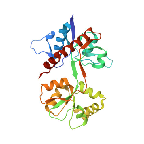
    Among the ionotropic glutamate receptors, the physiological role of kainate receptors is less well understood. Although ligands with selectivity toward the kainate receptor subtype GluK1 are available, tool compounds with selectivity at the remaining kainate receptor subtypes are sparse. Here, we have synthesized a series of quinoxaline-2,3-diones with substitutions in the N1-, 6-, and 7-position to investigate the structure-activity relationship (SAR) at GluK1-3 and GluK5. Pharmacological characterization at native and recombinant kainate and AMPA receptors revealed that compound 37 had a GluK3-binding affinity ( K i ) of 0.142 μM and 8-fold preference for GluK3 over GluK1. Despite lower binding affinity of 22 at GluK3 ( K i = 2.91 μM), its preference for GluK3 over GluK1 and GluK2 was >30-fold. Compound 37 was crystallized with the GluK1 ligand-binding domain to understand the SAR. The X-ray structure showed that 37 stabilized the protein in an open conformation, consistent with an antagonist binding mode.


  • Organizational Affiliation

    Department of Drug Design and Pharmacology, Faculty of Health and Medical Sciences , University of Copenhagen , DK-2100 Copenhagen , Denmark.


Macromolecules
Find similar proteins by:  (by identity cutoff)  |  3D Structure
Entity ID: 1
MoleculeChains Sequence LengthOrganismDetailsImage
Glutamate receptor ionotropic, kainate 1,Glutamate receptor ionotropic, kainate 1257Rattus norvegicusMutation(s): 1 
Gene Names: Grik1Glur5
UniProt
Find proteins for P22756 (Rattus norvegicus)
Explore P22756 
Go to UniProtKB:  P22756
Entity Groups  
Sequence Clusters30% Identity50% Identity70% Identity90% Identity95% Identity100% Identity
UniProt GroupP22756
Sequence Annotations
Expand
  • Reference Sequence
Small Molecules
Ligands 4 Unique
IDChains Name / Formula / InChI Key2D Diagram3D Interactions
EC8
Query on EC8

Download Ideal Coordinates CCD File 
H [auth A]N-(7-fluoro-2,3-dioxo-6-(trifluoromethyl)-3,4-dihydroquinoxalin-1(2H)-yl)-2-hydroxybenzamide
C16 H9 F4 N3 O4
MCKOVZZCGOMMIB-UHFFFAOYSA-N
SO4
Query on SO4

Download Ideal Coordinates CCD File 
G [auth A]SULFATE ION
O4 S
QAOWNCQODCNURD-UHFFFAOYSA-L
GOL
Query on GOL

Download Ideal Coordinates CCD File 
B [auth A],
C [auth A],
D [auth A],
E [auth A],
F [auth A]
GLYCEROL
C3 H8 O3
PEDCQBHIVMGVHV-UHFFFAOYSA-N
CL
Query on CL

Download Ideal Coordinates CCD File 
I [auth A]CHLORIDE ION
Cl
VEXZGXHMUGYJMC-UHFFFAOYSA-M
Binding Affinity Annotations 
IDSourceBinding Affinity
EC8 Binding MOAD:  6FZ4 Ki: 1120 (nM) from 1 assay(s)
Experimental Data & Validation

Experimental Data

  • Method: X-RAY DIFFRACTION
  • Resolution: 1.85 Å
  • R-Value Free: 0.216 
  • R-Value Work: 0.186 
  • R-Value Observed: 0.188 
  • Space Group: H 3 2
Unit Cell:
Length ( Å )Angle ( ˚ )
a = 88.867α = 90
b = 88.867β = 90
c = 157.145γ = 120
Software Package:
Software NamePurpose
SCALAdata scaling
PHASERphasing
PHENIXrefinement
PDB_EXTRACTdata extraction
XDSdata reduction

Structure Validation

View Full Validation Report


Ligand Structure Quality Assessment 


Entry History 

Deposition Data

Revision History  (Full details and data files)

  • Version 1.0: 2019-01-23
    Type: Initial release
  • Version 1.1: 2019-03-27
    Changes: Data collection, Database references
  • Version 1.2: 2024-01-17
    Changes: Data collection, Database references, Refinement description