6FNT

Ergothioneine-biosynthetic methyltransferase EgtD in complex with pyrrolidinohistidine


Experimental Data Snapshot

  • Method: X-RAY DIFFRACTION
  • Resolution: 1.90 Å
  • R-Value Free: 0.207 
  • R-Value Work: 0.170 
  • R-Value Observed: 0.172 

wwPDB Validation   3D Report Full Report


Ligand Structure Quality Assessment 


This is version 1.1 of the entry. See complete history


Literature

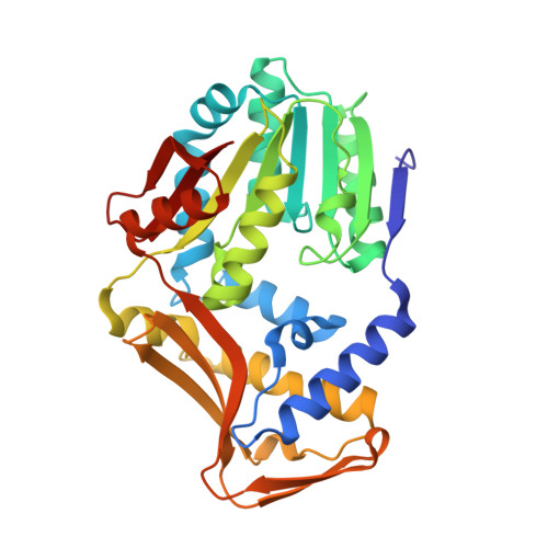
Inhibition and Regulation of the Ergothioneine Biosynthetic Methyltransferase EgtD.

Misson, L.Burn, R.Vit, A.Hildesheim, J.Beliaeva, M.A.Blankenfeldt, W.Seebeck, F.P.

(2018) ACS Chem Biol 13: 1333-1342

  • DOI: https://doi.org/10.1021/acschembio.8b00127
  • Primary Citation of Related Structures:  
    6FNQ, 6FNR, 6FNS, 6FNT

  • PubMed Abstract: 

    Ergothioneine is an emerging factor in cellular redox homeostasis in bacteria, fungi, plants, and animals. Reports that ergothioneine biosynthesis may be important for the pathogenicity of bacteria and fungi raise the question as to how this pathway is regulated and whether the corresponding enzymes may be therapeutic targets. The first step in ergothioneine biosynthesis is catalyzed by the methyltransferase EgtD that converts histidine into N-α-trimethylhistidine. This report examines the kinetic, thermodynamic and structural basis for substrate, product, and inhibitor binding by EgtD from Mycobacterium smegmatis. This study reveals an unprecedented substrate binding mechanism and a fine-tuned affinity landscape as determinants for product specificity and product inhibition. Both properties are evolved features that optimize the function of EgtD in the context of cellular ergothioneine production. On the basis of these findings, we developed a series of simple histidine derivatives that inhibit methyltransferase activity at low micromolar concentrations. Crystal structures of inhibited complexes validate this structure- and mechanism-based design strategy.


  • Organizational Affiliation

    Department for Chemistry , University of Basel , BPR 1096, Mattenstrasse 24a , Basel , Switzerland.


Macromolecules
Find similar proteins by:  (by identity cutoff)  |  3D Structure
Entity ID: 1
MoleculeChains Sequence LengthOrganismDetailsImage
Histidine N-alpha-methyltransferase
A, B
323Mycolicibacterium smegmatisMutation(s): 0 
Gene Names: egtDERS451418_06055
EC: 2.1.1.44
UniProt
Find proteins for A0R5M8 (Mycolicibacterium smegmatis (strain ATCC 700084 / mc(2)155))
Explore A0R5M8 
Go to UniProtKB:  A0R5M8
Entity Groups  
Sequence Clusters30% Identity50% Identity70% Identity90% Identity95% Identity100% Identity
UniProt GroupA0R5M8
Sequence Annotations
Expand
  • Reference Sequence
Small Molecules
Binding Affinity Annotations 
IDSourceBinding Affinity
AVO Binding MOAD:  6FNT Ki: 2.10e+4 (nM) from 1 assay(s)
Experimental Data & Validation

Experimental Data

  • Method: X-RAY DIFFRACTION
  • Resolution: 1.90 Å
  • R-Value Free: 0.207 
  • R-Value Work: 0.170 
  • R-Value Observed: 0.172 
  • Space Group: P 21 21 21
Unit Cell:
Length ( Å )Angle ( ˚ )
a = 71.768α = 90
b = 77.535β = 90
c = 139.045γ = 90
Software Package:
Software NamePurpose
XDSdata reduction
Aimlessdata scaling
PHENIXrefinement
PDB_EXTRACTdata extraction
PHENIXphasing

Structure Validation

View Full Validation Report


Ligand Structure Quality Assessment 


Entry History 

Deposition Data

Revision History  (Full details and data files)

  • Version 1.0: 2018-06-13
    Type: Initial release
  • Version 1.1: 2024-01-17
    Changes: Data collection, Database references, Refinement description