6FHW

Structure of Hormoconis resinae Glucoamylase


Experimental Data Snapshot

  • Method: X-RAY DIFFRACTION
  • Resolution: 3.60 Å
  • R-Value Free: 0.273 
  • R-Value Work: 0.264 
  • R-Value Observed: 0.265 

wwPDB Validation   3D Report Full Report


Ligand Structure Quality Assessment 


This is version 2.1 of the entry. See complete history


Literature

Structural insight into industrially relevant glucoamylases: flexible positions of starch-binding domains.

Roth, C.Moroz, O.V.Ariza, A.Skov, L.K.Ayabe, K.Davies, G.J.Wilson, K.S.

(2018) Acta Crystallogr D Struct Biol 74: 463-470

  • DOI: https://doi.org/10.1107/S2059798318004989
  • Primary Citation of Related Structures:  
    6FHV, 6FHW, 6FRV

  • PubMed Abstract: 

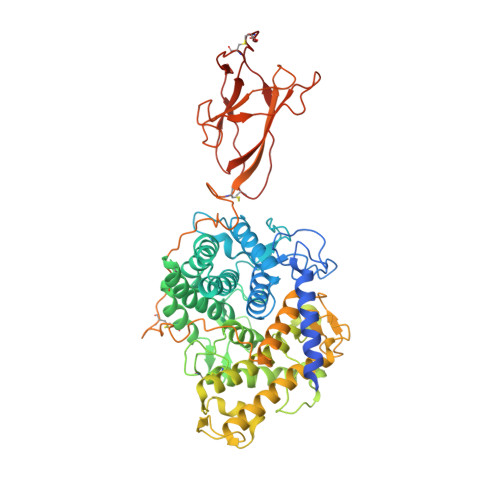
    Glucoamylases are one of the most important classes of enzymes in the industrial degradation of starch biomass. They consist of a catalytic domain and a carbohydrate-binding domain (CBM), with the latter being important for the interaction with the polymeric substrate. Whereas the catalytic mechanisms and structures of the individual domains are well known, the spatial arrangement of the domains with respect to each other and its influence on activity are not fully understood. Here, the structures of three industrially used fungal glucoamylases, two of which are full length, have been crystallized and determined. It is shown for the first time that the relative orientation between the CBM and the catalytic domain is flexible, as they can adopt different orientations independently of ligand binding, suggesting a role as an anchor to increase the contact time and the relative concentration of substrate near the active site. The flexibility in the orientations of the two domains presented a considerable challenge for the crystallization of the enzymes.


  • Organizational Affiliation

    Structural Biology Laboratory, Department of Chemistry, University of York, Heslington, York YO10 5DD, England.


Macromolecules
Find similar proteins by:  (by identity cutoff)  |  3D Structure
Entity ID: 1
MoleculeChains Sequence LengthOrganismDetailsImage
Glucoamylase P
A, B
616Amorphotheca resinaeMutation(s): 0 
Gene Names: GAMP
EC: 3.2.1.3
UniProt
Find proteins for Q03045 (Amorphotheca resinae)
Explore Q03045 
Go to UniProtKB:  Q03045
Entity Groups  
Sequence Clusters30% Identity50% Identity70% Identity90% Identity95% Identity100% Identity
UniProt GroupQ03045
Sequence Annotations
Expand
  • Reference Sequence
Oligosaccharides

Help

Entity ID: 2
MoleculeChains Length2D Diagram Glycosylation3D Interactions
2-acetamido-2-deoxy-beta-D-glucopyranose-(1-4)-2-acetamido-2-deoxy-beta-D-glucopyranose
C, D, E, G, H
C, D, E, G, H, I, J, K, L
2N-Glycosylation
Glycosylation Resources
GlyTouCan:  G42666HT
GlyCosmos:  G42666HT
GlyGen:  G42666HT
Entity ID: 3
MoleculeChains Length2D Diagram Glycosylation3D Interactions
alpha-D-mannopyranose-(1-6)-alpha-D-mannopyranose-(1-3)-beta-D-mannopyranose-(1-4)-2-acetamido-2-deoxy-beta-D-glucopyranose-(1-4)-2-acetamido-2-deoxy-beta-D-glucopyranose
F
5N-Glycosylation
Glycosylation Resources
GlyTouCan:  G77367MI
GlyCosmos:  G77367MI
GlyGen:  G77367MI
Entity ID: 4
MoleculeChains Length2D Diagram Glycosylation3D Interactions
4,6-dideoxy-4-{[(1S,4R,5S,6S)-4,5,6-trihydroxy-3-(hydroxymethyl)cyclohex-2-en-1-yl]amino}-alpha-D-glucopyranose-(1-4)-alpha-D-glucopyranose-(1-4)-alpha-D-glucopyranose
M, N
3N/AN/A
Glycosylation Resources
GlyTouCan:  G66431MI
GlyCosmos:  G66431MI
Small Molecules
Ligands 1 Unique
IDChains Name / Formula / InChI Key2D Diagram3D Interactions
NAG
Query on NAG

Download Ideal Coordinates CCD File 
O [auth A],
P [auth A]
2-acetamido-2-deoxy-beta-D-glucopyranose
C8 H15 N O6
OVRNDRQMDRJTHS-FMDGEEDCSA-N
Biologically Interesting Molecules (External Reference) 1 Unique
Experimental Data & Validation

Experimental Data

  • Method: X-RAY DIFFRACTION
  • Resolution: 3.60 Å
  • R-Value Free: 0.273 
  • R-Value Work: 0.264 
  • R-Value Observed: 0.265 
  • Space Group: P 21 21 21
Unit Cell:
Length ( Å )Angle ( ˚ )
a = 137.975α = 90
b = 149.827β = 90
c = 192.349γ = 90
Software Package:
Software NamePurpose
REFMACrefinement
PDB_EXTRACTdata extraction
XDSdata reduction
SCALAdata scaling
MOLREPphasing

Structure Validation

View Full Validation Report


Ligand Structure Quality Assessment 


Entry History 

Deposition Data

Revision History  (Full details and data files)

  • Version 1.0: 2018-05-09
    Type: Initial release
  • Version 2.0: 2020-07-29
    Type: Remediation
    Reason: Carbohydrate remediation
    Changes: Atomic model, Data collection, Derived calculations, Non-polymer description, Structure summary
  • Version 2.1: 2024-01-17
    Changes: Data collection, Database references, Derived calculations, Refinement description, Structure summary