6FH3

Protein arginine kinase McsB in the pArg-bound state


Experimental Data Snapshot

  • Method: X-RAY DIFFRACTION
  • Resolution: 1.85 Å
  • R-Value Free: 0.216 
  • R-Value Work: 0.181 
  • R-Value Observed: 0.182 

wwPDB Validation   3D Report Full Report


Ligand Structure Quality Assessment 


This is version 1.6 of the entry. See complete history


Literature

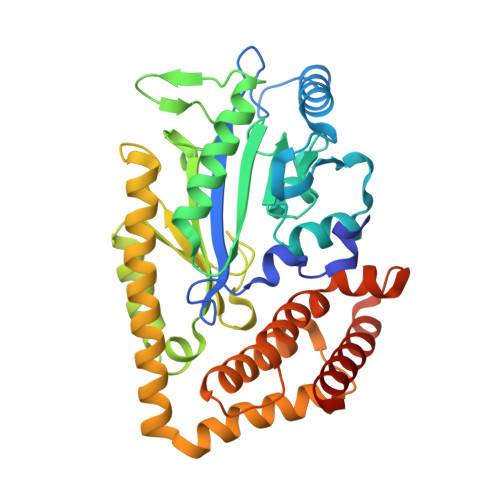
Structure of McsB, a protein kinase for regulated arginine phosphorylation.

Suskiewicz, M.J.Hajdusits, B.Beveridge, R.Heuck, A.Vu, L.D.Kurzbauer, R.Hauer, K.Thoeny, V.Rumpel, K.Mechtler, K.Meinhart, A.Clausen, T.

(2019) Nat Chem Biol 15: 510-518

  • DOI: https://doi.org/10.1038/s41589-019-0265-y
  • Primary Citation of Related Structures:  
    6FH1, 6FH2, 6FH3, 6FH4

  • PubMed Abstract: 

    Protein phosphorylation regulates key processes in all organisms. In Gram-positive bacteria, protein arginine phosphorylation plays a central role in protein quality control by regulating transcription factors and marking aberrant proteins for degradation. Here, we report structural, biochemical, and in vivo data of the responsible kinase, McsB, the founding member of an arginine-specific class of protein kinases. McsB differs in structure and mechanism from protein kinases that act on serine, threonine, and tyrosine residues and instead has a catalytic domain related to that of phosphagen kinases (PhKs), metabolic enzymes that phosphorylate small guanidino compounds. In McsB, the PhK-like phosphotransferase domain is structurally adapted to target protein substrates and is accompanied by a novel phosphoarginine (pArg)-binding domain that allosterically controls protein kinase activity. The identification of distinct pArg reader domains in this study points to a remarkably complex signaling system, thus challenging simplistic views of bacterial protein phosphorylation.


  • Organizational Affiliation

    Research Institute of Molecular Pathology (IMP), Vienna BioCenter (VBC), Vienna, Austria.


Macromolecules
Find similar proteins by:  (by identity cutoff)  |  3D Structure
Entity ID: 1
MoleculeChains Sequence LengthOrganismDetailsImage
Protein-arginine kinase
A, B
366Geobacillus stearothermophilusMutation(s): 0 
Gene Names: mcsB
EC: 2.7.14.1
UniProt
Find proteins for P0DMM5 (Geobacillus stearothermophilus)
Explore P0DMM5 
Go to UniProtKB:  P0DMM5
Entity Groups  
Sequence Clusters30% Identity50% Identity70% Identity90% Identity95% Identity100% Identity
UniProt GroupP0DMM5
Sequence Annotations
Expand
  • Reference Sequence
Small Molecules
Ligands 2 Unique
IDChains Name / Formula / InChI Key2D Diagram3D Interactions
RPI
Query on RPI

Download Ideal Coordinates CCD File 
C [auth A],
I [auth B]
phospho-arginine
C6 H15 N4 O5 P
CCTIOCVIZPCTGO-BYPYZUCNSA-N
EDO
Query on EDO

Download Ideal Coordinates CCD File 
D [auth A]
E [auth A]
F [auth A]
G [auth A]
H [auth A]
D [auth A],
E [auth A],
F [auth A],
G [auth A],
H [auth A],
J [auth B],
K [auth B],
L [auth B]
1,2-ETHANEDIOL
C2 H6 O2
LYCAIKOWRPUZTN-UHFFFAOYSA-N
Experimental Data & Validation

Experimental Data

  • Method: X-RAY DIFFRACTION
  • Resolution: 1.85 Å
  • R-Value Free: 0.216 
  • R-Value Work: 0.181 
  • R-Value Observed: 0.182 
  • Space Group: P 1 21 1
Unit Cell:
Length ( Å )Angle ( ˚ )
a = 49α = 90
b = 77.11β = 98.08
c = 94.13γ = 90
Software Package:
Software NamePurpose
PHENIXrefinement
XDSdata reduction
XSCALEdata scaling
PHASERphasing
Cootmodel building

Structure Validation

View Full Validation Report


Ligand Structure Quality Assessment 


Entry History 

Deposition Data

Revision History  (Full details and data files)

  • Version 1.0: 2019-02-27
    Type: Initial release
  • Version 1.1: 2019-04-10
    Changes: Data collection, Database references
  • Version 1.2: 2019-04-17
    Changes: Data collection, Database references
  • Version 1.3: 2019-04-24
    Changes: Data collection, Database references
  • Version 1.4: 2019-07-10
    Changes: Data collection
  • Version 1.5: 2019-07-17
    Changes: Data collection
  • Version 1.6: 2024-01-17
    Changes: Data collection, Database references, Refinement description