6FBP

Human Methionine Adenosyltransferase II mutant (S114A) in P22121 crystal form


Experimental Data Snapshot

  • Method: X-RAY DIFFRACTION
  • Resolution: 1.65 Å
  • R-Value Free: 0.195 
  • R-Value Work: 0.163 
  • R-Value Observed: 0.165 

wwPDB Validation   3D Report Full Report


Ligand Structure Quality Assessment 


This is version 1.2 of the entry. See complete history


Literature

Control and regulation of S-Adenosylmethionine biosynthesis by the regulatory beta subunit and quinolone-based compounds.

Panmanee, J.Bradley-Clarke, J.Mato, J.M.O'Neill, P.M.Antonyuk, S.V.Hasnain, S.S.

(2019) FEBS J 286: 2135-2154

  • DOI: https://doi.org/10.1111/febs.14790
  • Primary Citation of Related Structures:  
    6FBN, 6FBO, 6FBP, 6FCB, 6FCD, 6G6R

  • PubMed Abstract: 

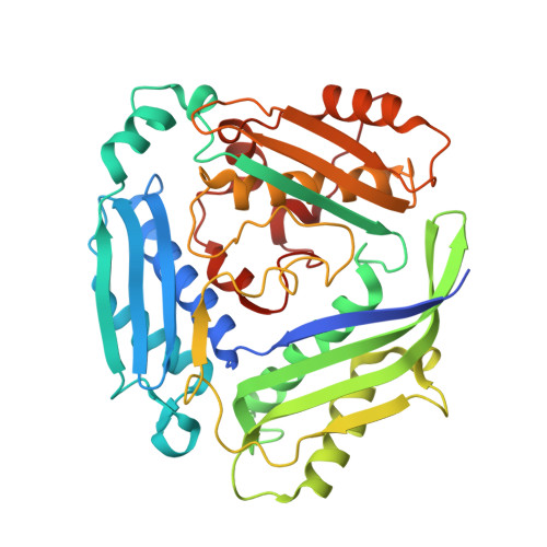
    Methylation is an underpinning process of life and provides control for biological processes such as DNA synthesis, cell growth, and apoptosis. Methionine adenosyltransferases (MAT) produce the cellular methyl donor, S-Adenosylmethionine (SAMe). Dysregulation of SAMe level is a relevant event in many diseases, including cancers such as hepatocellular carcinoma and colon cancer. In addition, mutation of Arg264 in MATα1 causes isolated persistent hypermethioninemia, which is characterized by low activity of the enzyme in liver and high level of plasma methionine. In mammals, MATα1/α2 and MATβV1/V2 are the catalytic and the major form of regulatory subunits, respectively. A gating loop comprising residues 113-131 is located beside the active site of catalytic subunits (MATα1/α2) and provides controlled access to the active site. Here, we provide evidence of how the gating loop facilitates the catalysis and define some of the key elements that control the catalytic efficiency. Mutation of several residues of MATα2 including Gln113, Ser114, and Arg264 lead to partial or total loss of enzymatic activity, demonstrating their critical role in catalysis. The enzymatic activity of the mutated enzymes is restored to varying degrees upon complex formation with MATβV1 or MATβV2, endorsing its role as an allosteric regulator of MATα2 in response to the levels of methionine or SAMe. Finally, the protein-protein interacting surface formed in MATα2:MATβ complexes is explored to demonstrate that several quinolone-based compounds modulate the activity of MATα2 and its mutants, providing a rational for chemical design/intervention responsive to the level of SAMe in the cellular environment. ENZYMES: Methionine adenosyltransferase (EC.2.5.1.6). DATABASE: Structural data are available in the RCSB PDB database under the PDB ID 6FBN (Q113A), 6FBP (S114A: P22 1 2 1 ), 6FBO (S114A: I222), 6FCB (P115G), 6FCD (R264A), 6FAJ (wtMATα2: apo), 6G6R (wtMATα2: holo).


  • Organizational Affiliation

    Molecular Biophysics Group, Institute of Integrative Biology, Faculty of Health and Life Sciences, University of Liverpool, UK.


Macromolecules
Find similar proteins by:  (by identity cutoff)  |  3D Structure
Entity ID: 1
MoleculeChains Sequence LengthOrganismDetailsImage
S-adenosylmethionine synthase isoform type-2
A, B
395Homo sapiensMutation(s): 0 
Gene Names: MAT2AAMS2MATA2
EC: 2.5.1.6
UniProt & NIH Common Fund Data Resources
Find proteins for P31153 (Homo sapiens)
Explore P31153 
Go to UniProtKB:  P31153
PHAROS:  P31153
GTEx:  ENSG00000168906 
Entity Groups  
Sequence Clusters30% Identity50% Identity70% Identity90% Identity95% Identity100% Identity
UniProt GroupP31153
Sequence Annotations
Expand
  • Reference Sequence
Small Molecules
Ligands 7 Unique
IDChains Name / Formula / InChI Key2D Diagram3D Interactions
ADN (Subject of Investigation/LOI)
Query on ADN

Download Ideal Coordinates CCD File 
M [auth B]ADENOSINE
C10 H13 N5 O4
OIRDTQYFTABQOQ-KQYNXXCUSA-N
PPK (Subject of Investigation/LOI)
Query on PPK

Download Ideal Coordinates CCD File 
J [auth B](DIPHOSPHONO)AMINOPHOSPHONIC ACID
H6 N O9 P3
PELPUMGXMYVGSQ-UHFFFAOYSA-N
PG4
Query on PG4

Download Ideal Coordinates CCD File 
C [auth A],
L [auth B]
TETRAETHYLENE GLYCOL
C8 H18 O5
UWHCKJMYHZGTIT-UHFFFAOYSA-N
EDO
Query on EDO

Download Ideal Coordinates CCD File 
E [auth A],
H [auth A],
I [auth A]
1,2-ETHANEDIOL
C2 H6 O2
LYCAIKOWRPUZTN-UHFFFAOYSA-N
ACT
Query on ACT

Download Ideal Coordinates CCD File 
D [auth A]ACETATE ION
C2 H3 O2
QTBSBXVTEAMEQO-UHFFFAOYSA-M
K (Subject of Investigation/LOI)
Query on K

Download Ideal Coordinates CCD File 
F [auth A],
N [auth B]
POTASSIUM ION
K
NPYPAHLBTDXSSS-UHFFFAOYSA-N
MG (Subject of Investigation/LOI)
Query on MG

Download Ideal Coordinates CCD File 
G [auth A],
K [auth B]
MAGNESIUM ION
Mg
JLVVSXFLKOJNIY-UHFFFAOYSA-N
Experimental Data & Validation

Experimental Data

  • Method: X-RAY DIFFRACTION
  • Resolution: 1.65 Å
  • R-Value Free: 0.195 
  • R-Value Work: 0.163 
  • R-Value Observed: 0.165 
  • Space Group: P 2 21 21
Unit Cell:
Length ( Å )Angle ( ˚ )
a = 62.09α = 90
b = 102.871β = 90
c = 108.17γ = 90
Software Package:
Software NamePurpose
REFMACrefinement
iMOSFLMdata reduction
Aimlessdata scaling
MOLREPphasing

Structure Validation

View Full Validation Report


Ligand Structure Quality Assessment 


Entry History 

Deposition Data

Revision History  (Full details and data files)

  • Version 1.0: 2019-01-30
    Type: Initial release
  • Version 1.1: 2019-08-14
    Changes: Data collection, Database references
  • Version 1.2: 2024-01-17
    Changes: Data collection, Database references, Derived calculations, Refinement description