6F9N

CRYSTAL STRUCTURE OF THE HUMAN CPSF160-WDR33 COMPLEX


Experimental Data Snapshot

  • Method: X-RAY DIFFRACTION
  • Resolution: 2.50 Å
  • R-Value Free: 0.263 
  • R-Value Work: 0.228 
  • R-Value Observed: 0.229 

wwPDB Validation   3D Report Full Report


This is version 1.1 of the entry. See complete history


Literature

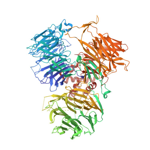
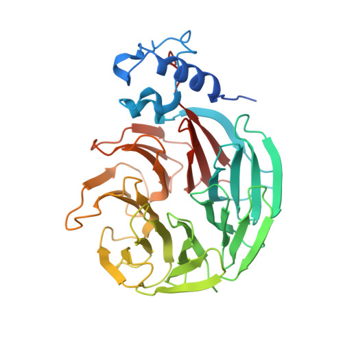
Structural insights into the assembly and polyA signal recognition mechanism of the human CPSF complex.

Clerici, M.Faini, M.Aebersold, R.Jinek, M.

(2017) Elife 6

  • DOI: https://doi.org/10.7554/eLife.33111
  • Primary Citation of Related Structures:  
    6F9N

  • PubMed Abstract: 

    3' polyadenylation is a key step in eukaryotic mRNA biogenesis. In mammalian cells, this process is dependent on the recognition of the hexanucleotide AAUAAA motif in the pre-mRNA polyadenylation signal by the cleavage and polyadenylation specificity factor (CPSF) complex. A core CPSF complex comprising CPSF160, WDR33, CPSF30 and Fip1 is sufficient for AAUAAA motif recognition, yet the molecular interactions underpinning its assembly and mechanism of PAS recognition are not understood. Based on cross-linking-coupled mass spectrometry, crystal structure of the CPSF160-WDR33 subcomplex and biochemical assays, we define the molecular architecture of the core human CPSF complex, identifying specific domains involved in inter-subunit interactions. In addition to zinc finger domains in CPSF30, we identify using quantitative RNA-binding assays an N-terminal lysine/arginine-rich motif in WDR33 as a critical determinant of specific AAUAAA motif recognition. Together, these results shed light on the function of CPSF in mediating PAS-dependent RNA cleavage and polyadenylation.


  • Organizational Affiliation

    Department of Biochemistry, University of Zurich, Zurich, Switzerland.


Macromolecules
Find similar proteins by:  (by identity cutoff)  |  3D Structure
Entity ID: 1
MoleculeChains Sequence LengthOrganismDetailsImage
Cleavage and polyadenylation specificity factor subunit 11,443Homo sapiensMutation(s): 0 
Gene Names: CPSF1CPSF160
UniProt & NIH Common Fund Data Resources
Find proteins for Q10570 (Homo sapiens)
Explore Q10570 
Go to UniProtKB:  Q10570
PHAROS:  Q10570
GTEx:  ENSG00000071894 
Entity Groups  
Sequence Clusters30% Identity50% Identity70% Identity90% Identity95% Identity100% Identity
UniProt GroupQ10570
Sequence Annotations
Expand
  • Reference Sequence
Find similar proteins by:  (by identity cutoff)  |  3D Structure
Entity ID: 2
MoleculeChains Sequence LengthOrganismDetailsImage
pre-mRNA 3' end processing protein WDR33379Homo sapiensMutation(s): 0 
Gene Names: WDR33WDC146
UniProt & NIH Common Fund Data Resources
Find proteins for Q9C0J8 (Homo sapiens)
Explore Q9C0J8 
Go to UniProtKB:  Q9C0J8
PHAROS:  Q9C0J8
GTEx:  ENSG00000136709 
Entity Groups  
Sequence Clusters30% Identity50% Identity70% Identity90% Identity95% Identity100% Identity
UniProt GroupQ9C0J8
Sequence Annotations
Expand
  • Reference Sequence
Experimental Data & Validation

Experimental Data

  • Method: X-RAY DIFFRACTION
  • Resolution: 2.50 Å
  • R-Value Free: 0.263 
  • R-Value Work: 0.228 
  • R-Value Observed: 0.229 
  • Space Group: P 1
Unit Cell:
Length ( Å )Angle ( ˚ )
a = 67.91α = 87.56
b = 77.4β = 76.41
c = 104.02γ = 67
Software Package:
Software NamePurpose
PHENIXrefinement
XDSdata reduction
XSCALEdata scaling
PHASERphasing
Cootmodel building

Structure Validation

View Full Validation Report


Entry History & Funding Information

Deposition Data


Funding OrganizationLocationGrant Number
European Research CouncilBelgiumERC-StG-337284

Revision History  (Full details and data files)

  • Version 1.0: 2018-01-03
    Type: Initial release
  • Version 1.1: 2024-01-17
    Changes: Data collection, Database references, Refinement description