The ancestral retinoic acid receptor was a low-affinity sensor triggering neuronal differentiation.
Handberg-Thorsager, M., Gutierrez-Mazariegos, J., Arold, S.T., Kumar Nadendla, E., Bertucci, P.Y., Germain, P., Tomancak, P., Pierzchalski, K., Jones, J.W., Albalat, R., Kane, M.A., Bourguet, W., Laudet, V., Arendt, D., Schubert, M.(2018) Sci Adv 4: eaao1261-eaao1261
- PubMed: 29492455
- DOI: https://doi.org/10.1126/sciadv.aao1261
- Primary Citation of Related Structures:
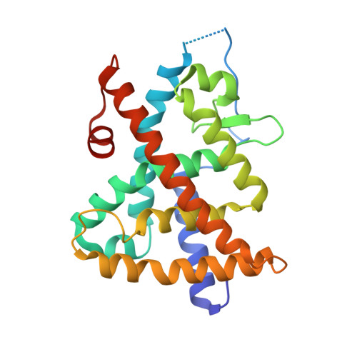
6EU9 - PubMed Abstract:
Retinoic acid (RA) is an important intercellular signaling molecule in vertebrate development, with a well-established role in the regulation of hox genes during hindbrain patterning and in neurogenesis. However, the evolutionary origin of the RA signaling pathway remains elusive. To elucidate the evolution of the RA signaling system, we characterized RA metabolism and signaling in the marine annelid Platynereis dumerilii , a powerful model for evolution, development, and neurobiology. Binding assays and crystal structure analyses show that the annelid retinoic acid receptor (RAR) binds RA and activates transcription just as vertebrate RARs, yet with a different ligand-binding pocket and lower binding affinity, suggesting a permissive rather than instructive role of RA signaling. RAR knockdown and RA treatment of swimming annelid larvae further reveal that the RA signal is locally received in the medial neuroectoderm, where it controls neurogenesis and axon outgrowth, whereas the spatial colinear hox gene expression in the neuroectoderm remains unaffected. These findings suggest that one early role of the new RAR in bilaterian evolution was to control the spatially restricted onset of motor and interneuron differentiation in the developing ventral nerve cord and to indicate that the regulation of hox -controlled anterior-posterior patterning arose only at the base of the chordates, concomitant with a high-affinity RAR needed for the interpretation of a complex RA gradient.
Organizational Affiliation:
Developmental Biology Unit, European Molecular Biology Laboratory, Meyerhofstrasse 1, 69012 Heidelberg, Germany.