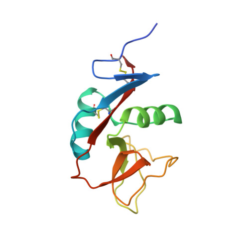
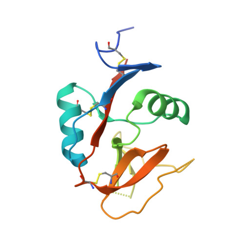
6E7D

Structure of the inhibitory NKR-P1B receptor bound to the host-encoded ligand, Clr-b


Experimental Data Snapshot

  • Method: X-RAY DIFFRACTION
  • Resolution: 2.90 Å
  • R-Value Free: 0.225 
  • R-Value Work: 0.186 
  • R-Value Observed: 0.188 

wwPDB Validation   3D Report Full Report


Ligand Structure Quality Assessment 


This is version 1.4 of the entry. See complete history


Literature

Recognition of host Clr-b by the inhibitory NKR-P1B receptor provides a basis for missing-self recognition.

Balaji, G.R.Aguilar, O.A.Tanaka, M.Shingu-Vazquez, M.A.Fu, Z.Gully, B.S.Lanier, L.L.Carlyle, J.R.Rossjohn, J.Berry, R.

(2018) Nat Commun 9: 4623-4623

  • DOI: https://doi.org/10.1038/s41467-018-06989-2
  • Primary Citation of Related Structures:  
    6E7D

  • PubMed Abstract: 

    The interaction between natural killer (NK) cell inhibitory receptors and their cognate ligands constitutes a key mechanism by which healthy tissues are protected from NK cell-mediated lysis. However, self-ligand recognition remains poorly understood within the prototypical NKR-P1 receptor family. Here we report the structure of the inhibitory NKR-P1B receptor bound to its cognate host ligand, Clr-b. NKR-P1B and Clr-b interact via a head-to-head docking mode through an interface that includes a large array of polar interactions. NKR-P1B:Clr-b recognition is extremely sensitive to mutations at the heterodimeric interface, with most mutations severely impacting both Clr-b binding and NKR-P1B receptor function to implicate a low affinity interaction. Within the structure, two NKR-P1B:Clr-b complexes are cross-linked by a non-classic NKR-P1B homodimer, and the disruption of homodimer formation abrogates Clr-b recognition. These data provide an insight into a fundamental missing-self recognition system and suggest an avidity-based mechanism underpins NKR-P1B receptor function.


  • Organizational Affiliation

    Infection and Immunity Program and Department of Biochemistry and Molecular Biology, Biomedicine Discovery Institute, Monash University, Clayton, Victoria, 3800, Australia.


Macromolecules
Find similar proteins by:  (by identity cutoff)  |  3D Structure
Entity ID: 1
MoleculeChains Sequence LengthOrganismDetailsImage
C-type lectin domain family 2 member D
A, B, C, D, E
A, B, C, D, E, F, G, H, I, J, K, L, M, N, O, P
124Mus musculusMutation(s): 0 
Gene Names: Clec2dClrbOcil
UniProt
Find proteins for Q91V08 (Mus musculus)
Explore Q91V08 
Go to UniProtKB:  Q91V08
Entity Groups  
Sequence Clusters30% Identity50% Identity70% Identity90% Identity95% Identity100% Identity
UniProt GroupQ91V08
Sequence Annotations
Expand
  • Reference Sequence
Find similar proteins by:  (by identity cutoff)  |  3D Structure
Entity ID: 2
MoleculeChains Sequence LengthOrganismDetailsImage
Killer cell lectin-like receptor subfamily B member 1B allele B
Q, R, S, T, U
Q, R, S, T, U, V, W, X
137Mus musculusMutation(s): 1 
Gene Names: Klrb1bKlrb1dLy55dNkrp1bNkrp1d
UniProt
Find proteins for Q99JB4 (Mus musculus)
Explore Q99JB4 
Go to UniProtKB:  Q99JB4
Entity Groups  
Sequence Clusters30% Identity50% Identity70% Identity90% Identity95% Identity100% Identity
UniProt GroupQ99JB4
Sequence Annotations
Expand
  • Reference Sequence
Small Molecules
Ligands 2 Unique
IDChains Name / Formula / InChI Key2D Diagram3D Interactions
NAG
Query on NAG

Download Ideal Coordinates CCD File 
BA [auth R]
DA [auth S]
FA [auth T]
HA [auth U]
JA [auth V]
BA [auth R],
DA [auth S],
FA [auth T],
HA [auth U],
JA [auth V],
LA [auth W],
NA [auth X],
Z [auth Q]
2-acetamido-2-deoxy-beta-D-glucopyranose
C8 H15 N O6
OVRNDRQMDRJTHS-FMDGEEDCSA-N
SO4
Query on SO4

Download Ideal Coordinates CCD File 
AA [auth Q]
CA [auth R]
EA [auth S]
GA [auth T]
IA [auth U]
AA [auth Q],
CA [auth R],
EA [auth S],
GA [auth T],
IA [auth U],
KA [auth V],
MA [auth W],
OA [auth X],
Y [auth D]
SULFATE ION
O4 S
QAOWNCQODCNURD-UHFFFAOYSA-L
Experimental Data & Validation

Experimental Data

  • Method: X-RAY DIFFRACTION
  • Resolution: 2.90 Å
  • R-Value Free: 0.225 
  • R-Value Work: 0.186 
  • R-Value Observed: 0.188 
  • Space Group: P 1
Unit Cell:
Length ( Å )Angle ( ˚ )
a = 66.93α = 73.12
b = 122.069β = 82.08
c = 131.561γ = 84.46
Software Package:
Software NamePurpose
BUSTERrefinement
iMOSFLMdata reduction
SCALAdata scaling
MOLREPphasing

Structure Validation

View Full Validation Report


Ligand Structure Quality Assessment 


Entry History & Funding Information

Deposition Data


Funding OrganizationLocationGrant Number
National Health and Medical Research Council (NHMRC, Australia)AustraliaAPP1109901
Australian Research Council (ARC)AustraliaFL160100049

Revision History  (Full details and data files)

  • Version 1.0: 2018-10-24
    Type: Initial release
  • Version 1.1: 2019-05-08
    Changes: Data collection, Database references
  • Version 1.2: 2020-01-01
    Changes: Author supporting evidence
  • Version 1.3: 2020-07-29
    Type: Remediation
    Reason: Carbohydrate remediation
    Changes: Data collection, Derived calculations, Structure summary
  • Version 1.4: 2023-10-11
    Changes: Data collection, Database references, Refinement description, Structure summary