6E4V

The Crystal Structure of FhuE from E. coli in complex with its substrate Coprogen


Experimental Data Snapshot

  • Method: X-RAY DIFFRACTION
  • Resolution: 2.00 Å
  • R-Value Free: 0.235 
  • R-Value Work: 0.206 
  • R-Value Observed: 0.208 

wwPDB Validation   3D Report Full Report


Ligand Structure Quality Assessment 


This is version 1.3 of the entry. See complete history


Literature

Determination of the molecular basis for coprogen import by Gram-negative bacteria.

Grinter, R.Lithgow, T.

(2019) IUCrJ 6: 401-411

  • DOI: https://doi.org/10.1107/S2052252519002926
  • Primary Citation of Related Structures:  
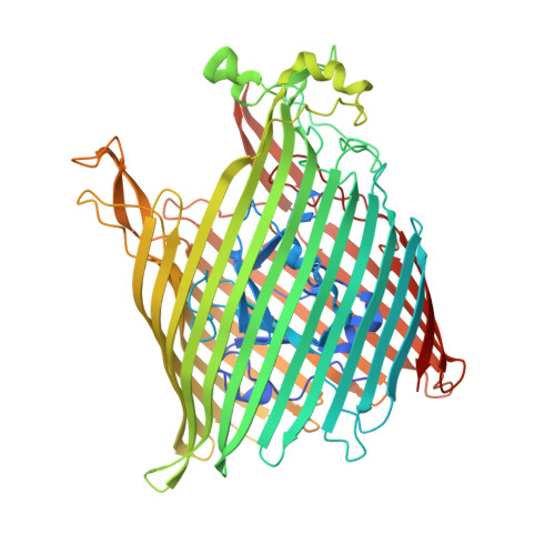
    6E4V

  • PubMed Abstract: 

    In order to survive in mixed microbial communities, some species of fungi secrete coprogens, siderophores that facilitate capture of the scarce nutrient iron. The TonB-dependent transporter FhuE is integrated in the outer membrane of Gram-negative bacteria and has been reported to scavenge these fungally produced coprogens. In this work, an Escherichia coli strain was engineered that is dependent solely on FhuE for its access to siderophore-sequestered iron. Using this tool, it is shown that while FhuE is highly active in the import of coprogens, it has some level of promiscuity, acting as a low-affinity transporter for related siderophores. The crystal structure of FhuE in complex with coprogen was determined, providing a structural basis to explain this selective promiscuity. The structural data, in combination with functional analysis, presented in this work show that FhuE has evolved to specifically engage with planar siderophores. A potential evolutionary driver, and a critical consequence of this selectivity, is that it allows FhuE to exclude antibiotics that mimic nonplanar hydroxamate siderophores: these toxic molecules could otherwise cross the outer membrane barrier through a Trojan horse mechanism.


  • Organizational Affiliation

    Infection and Immunity Program, Biomedicine Discovery Institute and Department of Microbiology, Monash University, Monash, Victoria 3800, Australia.


Macromolecules
Find similar proteins by:  (by identity cutoff)  |  3D Structure
Entity ID: 1
MoleculeChains Sequence LengthOrganismDetailsImage
FhuE receptor693Escherichia coli BW25113Mutation(s): 0 
Gene Names: fhuEb1102JW1088
Membrane Entity: Yes 
UniProt
Find proteins for P16869 (Escherichia coli (strain K12))
Explore P16869 
Go to UniProtKB:  P16869
Entity Groups  
Sequence Clusters30% Identity50% Identity70% Identity90% Identity95% Identity100% Identity
UniProt GroupP16869
Sequence Annotations
Expand
  • Reference Sequence
Experimental Data & Validation

Experimental Data

  • Method: X-RAY DIFFRACTION
  • Resolution: 2.00 Å
  • R-Value Free: 0.235 
  • R-Value Work: 0.206 
  • R-Value Observed: 0.208 
  • Space Group: P 21 21 21
Unit Cell:
Length ( Å )Angle ( ˚ )
a = 68.19α = 90
b = 103.86β = 90
c = 118.62γ = 90
Software Package:
Software NamePurpose
BUSTERrefinement
XDSdata reduction
Aimlessdata scaling
PHENIXphasing

Structure Validation

View Full Validation Report


Ligand Structure Quality Assessment 


Entry History & Funding Information

Deposition Data


Funding OrganizationLocationGrant Number
Wellcome TrustUnited Kingdom106077/Z/14/Z

Revision History  (Full details and data files)

  • Version 1.0: 2019-04-10
    Type: Initial release
  • Version 1.1: 2019-05-29
    Changes: Data collection, Database references
  • Version 1.2: 2020-07-29
    Type: Remediation
    Reason: Carbohydrate remediation
    Changes: Data collection, Derived calculations, Refinement description, Structure summary
  • Version 1.3: 2023-10-11
    Changes: Data collection, Database references, Refinement description, Structure summary