6E2V

MDDEF in complex with MVAPP, ADPBeF3 and magnesium


Experimental Data Snapshot

  • Method: X-RAY DIFFRACTION
  • Resolution: 2.10 Å
  • R-Value Free: 0.193 
  • R-Value Work: 0.174 
  • R-Value Observed: 0.175 

wwPDB Validation   3D Report Full Report


Ligand Structure Quality Assessment 


This is version 1.2 of the entry. See complete history


Literature

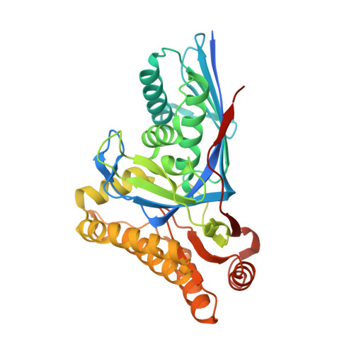
Visualizing the enzyme mechanism of mevalonate diphosphate decarboxylase.

Chen, C.L.Paul, L.N.Mermoud, J.C.Steussy, C.N.Stauffacher, C.V.

(2020) Nat Commun 11: 3969-3969

  • DOI: https://doi.org/10.1038/s41467-020-17733-0
  • Primary Citation of Related Structures:  
    6E2S, 6E2T, 6E2U, 6E2V, 6E2W, 6E2Y

  • PubMed Abstract: 

    Mevalonate diphosphate decarboxylases (MDDs) catalyze the ATP-dependent-Mg 2+ -decarboxylation of mevalonate-5-diphosphate (MVAPP) to produce isopentenyl diphosphate (IPP), which is essential in both eukaryotes and prokaryotes for polyisoprenoid synthesis. The substrates, MVAPP and ATP, have been shown to bind sequentially to MDD. Here we report crystals in which the enzyme remains active, allowing the visualization of conformational changes in Enterococcus faecalis MDD that describe sequential steps in an induced fit enzymatic reaction. Initial binding of MVAPP modulates the ATP binding pocket with a large loop movement. Upon ATP binding, a phosphate binding loop bends over the active site to recognize ATP and bring the molecules to their catalytically favored configuration. Positioned substrates then can chelate two Mg 2+ ions for the two steps of the reaction. Closure of the active site entrance brings a conserved lysine to trigger dissociative phosphoryl transfer of γ-phosphate from ATP to MVAPP, followed by the production of IPP.


  • Organizational Affiliation

    Department of Biological Sciences, Purdue University, West Lafayette, IN, 47907, USA.


Macromolecules
Find similar proteins by:  (by identity cutoff)  |  3D Structure
Entity ID: 1
MoleculeChains Sequence LengthOrganismDetailsImage
Mevalonate diphosphate decarboxylase355Enterococcus faecalisMutation(s): 0 
Gene Names: mvaDB6S42_02310
EC: 4.1.1.33
UniProt
Find proteins for Q9FD68 (Enterococcus faecalis)
Explore Q9FD68 
Go to UniProtKB:  Q9FD68
Entity Groups  
Sequence Clusters30% Identity50% Identity70% Identity90% Identity95% Identity100% Identity
UniProt GroupQ9FD68
Sequence Annotations
Expand
  • Reference Sequence
Small Molecules
Ligands 4 Unique
IDChains Name / Formula / InChI Key2D Diagram3D Interactions
ADP (Subject of Investigation/LOI)
Query on ADP

Download Ideal Coordinates CCD File 
F [auth A]ADENOSINE-5'-DIPHOSPHATE
C10 H15 N5 O10 P2
XTWYTFMLZFPYCI-KQYNXXCUSA-N
DP6 (Subject of Investigation/LOI)
Query on DP6

Download Ideal Coordinates CCD File 
D [auth A](3R)-3-HYDROXY-5-{[(R)-HYDROXY(PHOSPHONOOXY)PHOSPHORYL]OXY}-3-METHYLPENTANOIC ACID
C6 H14 O10 P2
SIGQQUBJQXSAMW-ZCFIWIBFSA-N
BEF (Subject of Investigation/LOI)
Query on BEF

Download Ideal Coordinates CCD File 
E [auth A]BERYLLIUM TRIFLUORIDE ION
Be F3
OGIAHMCCNXDTIE-UHFFFAOYSA-K
MG (Subject of Investigation/LOI)
Query on MG

Download Ideal Coordinates CCD File 
B [auth A],
C [auth A]
MAGNESIUM ION
Mg
JLVVSXFLKOJNIY-UHFFFAOYSA-N
Experimental Data & Validation

Experimental Data

  • Method: X-RAY DIFFRACTION
  • Resolution: 2.10 Å
  • R-Value Free: 0.193 
  • R-Value Work: 0.174 
  • R-Value Observed: 0.175 
  • Space Group: P 21 21 2
Unit Cell:
Length ( Å )Angle ( ˚ )
a = 79.636α = 90
b = 98.599β = 90
c = 45.632γ = 90
Software Package:
Software NamePurpose
PHENIXrefinement
HKL-2000data reduction
HKL-2000data scaling
PHASERphasing

Structure Validation

View Full Validation Report


Ligand Structure Quality Assessment 


Entry History 

Deposition Data

Revision History  (Full details and data files)

  • Version 1.0: 2020-01-15
    Type: Initial release
  • Version 1.1: 2020-08-19
    Changes: Database references, Derived calculations
  • Version 1.2: 2023-10-11
    Changes: Data collection, Database references, Refinement description