


Crystal structure of the Acinetobacter phage vB_ApiP_P1 tailspike protein
Plattner, M., Shneider, M.M., Oliveira, H., Azeredo, J., Leiman, P.G.To be published.
Experimental Data Snapshot
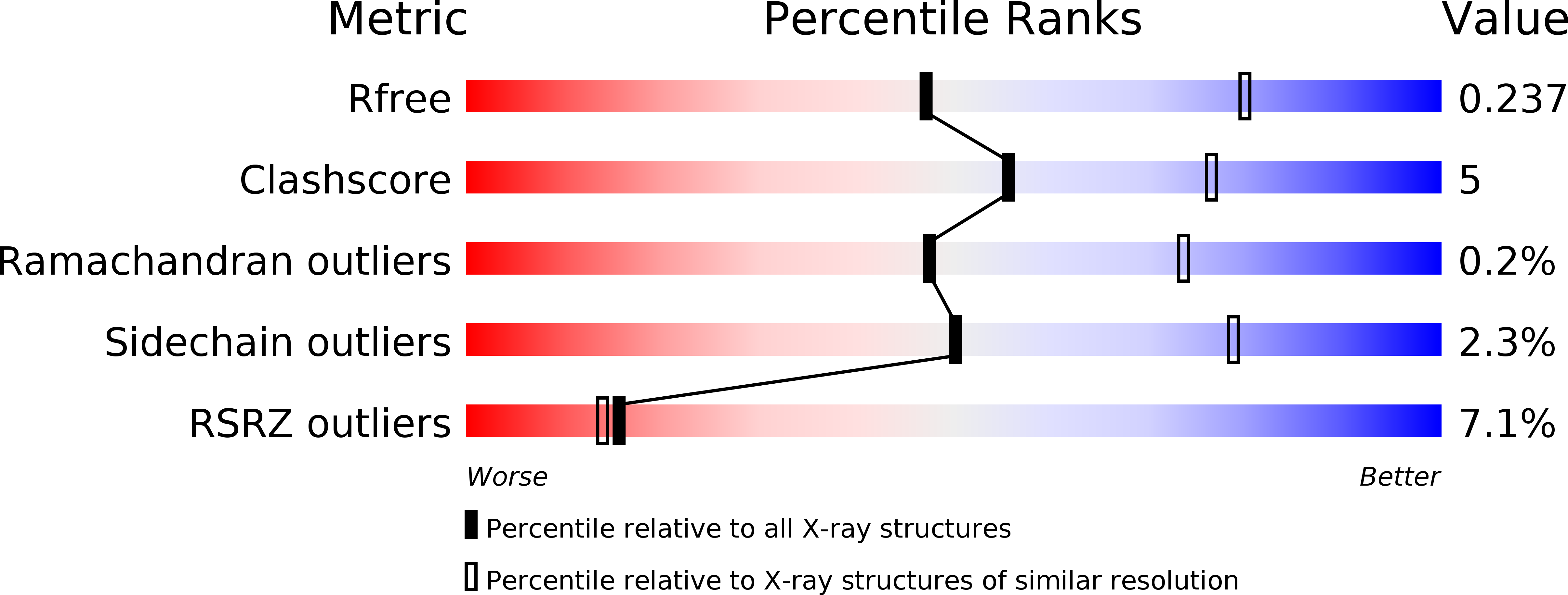
wwPDB Validation 3D Report Full Report
Entity ID: 1 | |||||
|---|---|---|---|---|---|
| Molecule | Chains | Sequence Length | Organism | Details | Image |
| Tailspike protein | 554 | Acinetobacter phage vB_ApiP_P1 | Mutation(s): 0 Gene Names: P1_43 | 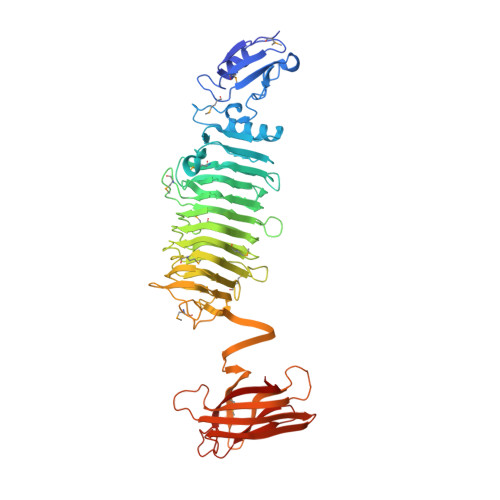 | |
UniProt | |||||
Find proteins for A0A221SBY4 (Acinetobacter phage vB_ApiP_P1) Explore A0A221SBY4 Go to UniProtKB: A0A221SBY4 | |||||
Entity Groups | |||||
| Sequence Clusters | 30% Identity50% Identity70% Identity90% Identity95% Identity100% Identity | ||||
| UniProt Group | A0A221SBY4 | ||||
Sequence AnnotationsExpand | |||||
| |||||
| Ligands 2 Unique | |||||
|---|---|---|---|---|---|
| ID | Chains | Name / Formula / InChI Key | 2D Diagram | 3D Interactions | |
| CL Query on CL | G [auth A], J [auth C], L [auth F] | CHLORIDE ION Cl VEXZGXHMUGYJMC-UHFFFAOYSA-M |  | ||
| NA Query on NA | H [auth B], I [auth C], K [auth D] | SODIUM ION Na FKNQFGJONOIPTF-UHFFFAOYSA-N |  | ||
| Modified Residues 1 Unique | |||||
|---|---|---|---|---|---|
| ID | Chains | Type | Formula | 2D Diagram | Parent |
| MSE Query on MSE | A, B, C, D, E A, B, C, D, E, F | L-PEPTIDE LINKING | C5 H11 N O2 Se |  | MET |
| Length ( Å ) | Angle ( ˚ ) |
|---|---|
| a = 81.123 | α = 90 |
| b = 90.015 | β = 90 |
| c = 508.604 | γ = 90 |
| Software Name | Purpose |
|---|---|
| PHENIX | refinement |
| XDS | data reduction |
| SCALA | data scaling |
| HKL2Map | phasing |
RCSB PDB (citation) is hosted by
RCSB PDB is a member of the

