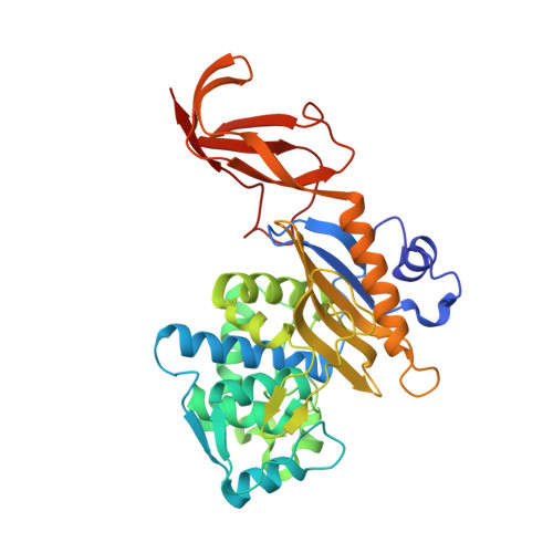
Recognition of Peptidoglycan Fragments by the Transpeptidase PBP4 FromStaphylococcus aureus.
Maya-Martinez, R., Alexander, J.A.N., Otten, C.F., Ayala, I., Vollmer, D., Gray, J., Bougault, C.M., Burt, A., Laguri, C., Fonvielle, M., Arthur, M., Strynadka, N.C.J., Vollmer, W., Simorre, J.P.(2018) Front Microbiol 9: 3223-3223
- PubMed: 30713527
- DOI: https://doi.org/10.3389/fmicb.2018.03223
- Primary Citation of Related Structures:
6DZ8 - PubMed Abstract:
Peptidoglycan (PG) is an essential component of the cell envelope, maintaining bacterial cell shape and protecting it from bursting due to turgor pressure. The monoderm bacterium Staphylococcus aureus has a highly cross-linked PG, with ~90% of peptide stems participating in DD-cross-links and up to 15 peptide stems connected with each other. These cross-links are formed in transpeptidation reactions catalyzed by penicillin-binding proteins (PBPs) of classes A and B. Most S. aureus strains have three housekeeping PBPs with this function (PBP1, PBP2, and PBP3) but MRSA strains have acquired a third class B PBP, PBP2a, which is encoded by the mecA gene and required for the expression of high-level resistance to β-lactams. Another housekeeping PBP of S. aureus is PBP4, which belongs to the class C PBPs, and hence would be expected to have PG hydrolase (DD-carboxypeptidase or DD-endopeptidase) activity. However, previous works showed that, unexpectedly, PBP4 has transpeptidase activity that significantly contributes to both the high level of cross-linking in the PG of S. aureus and to the low level of β-lactam resistance in the absence of PBP2a. To gain insights into this unusual activity of PBP4, we studied by NMR spectroscopy its interaction in vitro with different substrates, including intact peptidoglycan, synthetic peptide stems, muropeptides, and long glycan chains with uncross-linked peptide stems. PBP4 showed no affinity for the complex, intact peptidoglycan or the smallest isolated peptide stems. Transpeptidase activity of PBP4 was verified with the disaccharide peptide subunits (muropeptides) in vitro , producing cyclic dimer and multimer products; these assays also showed a designed PBP4(S75C) nucleophile mutant to be inactive. Using this inactive but structurally highly similar variant, liquid-state NMR identified two interaction surfaces in close proximity to the central nucleophile position that can accommodate the potential donor and acceptor stems for the transpeptidation reaction. A PBP4:muropeptide model structure was built from these experimental restraints, which provides new mechanistic insights into mecA independent resistance to β-lactams in S. aureus .
Organizational Affiliation:
University Grenoble Alpes, CNRS, CEA, IBS, Grenoble, France.