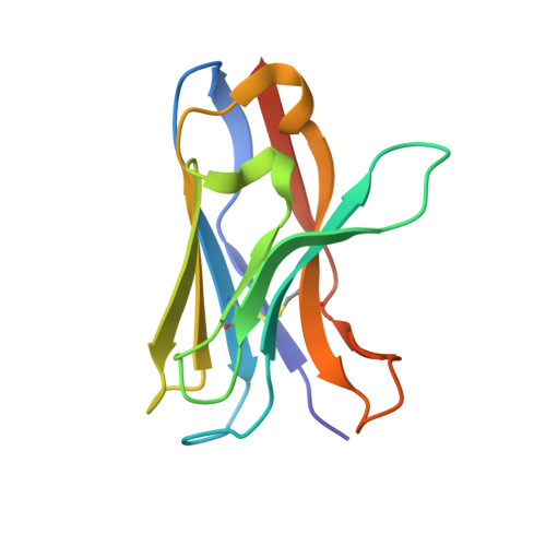
Structure of a VHH isolated from a naive phage display library.
White, B., Huh, I., Brooks, C.L.(2019) BMC Res Notes 12: 154-154
- PubMed: 30890176
- DOI: https://doi.org/10.1186/s13104-019-4197-0
- Primary Citation of Related Structures:
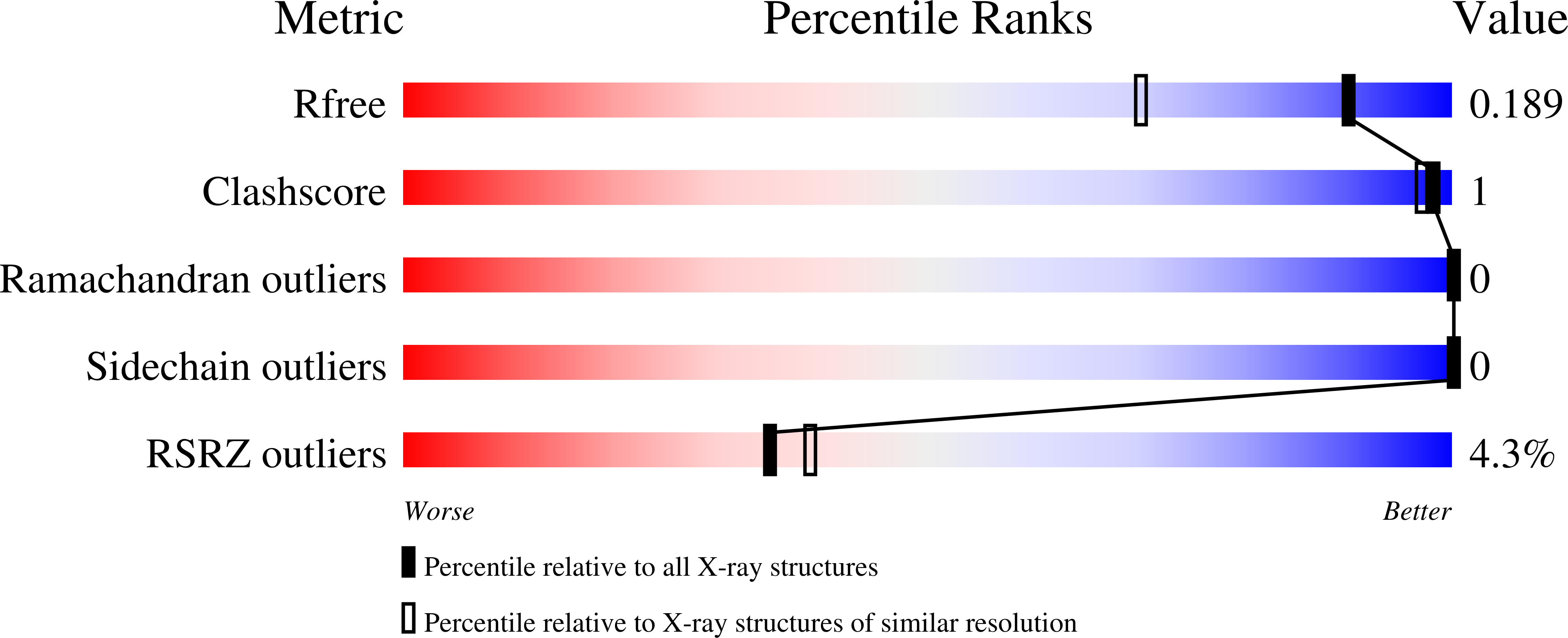
6DYX - PubMed Abstract:
To determine the X-ray structure and biophysical properties of a Camelid V H H isolated from a naïve phage display library.
Organizational Affiliation:
Department of Chemistry, California State University Fresno, 2555 E San Ramon Ave, Fresno, CA, 93740, USA.